[English] 日本語
Yorodumi
- PDB-7fqj: Crystal Structure of human Legumain in complex with (2S)-N-[(1S)-... -

+
Open data


ID or keywords:

Loading...

-
Basic information

Entry
Database: PDB / ID: 7fqj
TitleCrystal Structure of human Legumain in complex with (2S)-N-[(1S)-3-amino-1-cyano-3-oxopropyl]-1-[1-[4-[(2,4-difluorophenyl)methoxy]phenyl]cyclopropanecarbonyl]pyrrolidine-2-carboxamide
ComponentsLegumain
KeywordsHYDROLASE/INHIBITOR / CYSTEINE PROTEASE / ALLOSTERIC INHIBITOR / ASPARAGINYL ENDOPEPTIDASE / ALZHEIMER'S DISEASE / HYDROLASE / HYDROLASE-INHIBITOR complex
Function / homology
Function and homology information


negative regulation of ERBB signaling pathway / legumain / vacuolar protein processing / renal system process / Vitamin D (calciferol) metabolism / receptor catabolic process / vitamin D metabolic process / self proteolysis / endolysosome lumen / response to acidic pH ...negative regulation of ERBB signaling pathway / legumain / vacuolar protein processing / renal system process / Vitamin D (calciferol) metabolism / receptor catabolic process / vitamin D metabolic process / self proteolysis / endolysosome lumen / response to acidic pH / activation of cysteine-type endopeptidase activity / dendritic spine organization / positive regulation of monocyte chemotaxis / Trafficking and processing of endosomal TLR / positive regulation of endothelial cell chemotaxis / negative regulation of multicellular organism growth / cellular response to hepatocyte growth factor stimulus / associative learning / protein maturation / endopeptidase activator activity / cellular response to calcium ion / MHC class II antigen presentation / positive regulation of mitotic cell cycle / lysosomal lumen / proteolysis involved in protein catabolic process / positive regulation of long-term synaptic potentiation / tau protein binding / memory / cellular response to amyloid-beta / antigen processing and presentation of exogenous peptide antigen via MHC class II / late endosome / apical part of cell / peptidase activity / negative regulation of neuron apoptotic process / lysosome / cysteine-type endopeptidase activity / negative regulation of gene expression / positive regulation of cell population proliferation / perinuclear region of cytoplasm / proteolysis / extracellular exosome / extracellular region / cytoplasm
Similarity search - Function
: / Legumain, prodomain / Legumain prodomain superfamily / Asparaginyl endopeptidase / Peptidase C13, legumain / Peptidase C13 family
Similarity search - Domain/homology
CITRATE ANION / Chem-WSN / Legumain
Similarity search - Component
Biological speciesHomo sapiens (human)
MethodX-RAY DIFFRACTION / SYNCHROTRON / MOLECULAR REPLACEMENT / Resolution: 1.7 Å
AuthorsEhler, A. / Benz, J. / Bartels, B. / Rudolph, M.G.
Funding support Switzerland, 1items
OrganizationGrant numberCountry
F. Hoffmann-La Roche Switzerland
CitationJournal: To be published
Title: Crystal Structure of a human Legumain complex
Authors: Bartels, B. / Kuhn, B. / Benz, J. / Rudolph, M.G.
History
DepositionOct 5, 2022Deposition site: RCSB / Processing site: RCSB
Revision 1.0Oct 11, 2023Provider: repository / Type: Initial release
Revision 2.0Nov 15, 2023Group: Atomic model / Data collection / Derived calculations
Category: atom_site / chem_comp_atom ...atom_site / chem_comp_atom / chem_comp_bond / pdbx_validate_rmsd_angle / pdbx_validate_torsion / struct_conn
Item: _atom_site.auth_atom_id / _atom_site.label_atom_id ..._atom_site.auth_atom_id / _atom_site.label_atom_id / _chem_comp_atom.atom_id / _chem_comp_bond.atom_id_1 / _chem_comp_bond.atom_id_2 / _struct_conn.pdbx_leaving_atom_flag / _struct_conn.ptnr1_label_atom_id / _struct_conn.ptnr2_label_atom_id

-
Structure visualization

Structure viewerMolecule:
MolmilJmol/JSmol

Downloads & links

-
Assembly

Deposited unit
A: Legumain
B: Legumain
C: Legumain
hetero molecules


Theoretical massNumber of molelcules
Total (without water)155,98618
Polymers152,6333
Non-polymers3,35315
Water11,962664
1
A: Legumain
hetero molecules


Theoretical massNumber of molelcules
Total (without water)52,0926
Polymers50,8781
Non-polymers1,2145
Water181
TypeNameSymmetry operationNumber
identity operation1_555x,y,z1
2
B: Legumain
hetero molecules


Theoretical massNumber of molelcules
Total (without water)52,0145
Polymers50,8781
Non-polymers1,1364
Water181
TypeNameSymmetry operationNumber
identity operation1_555x,y,z1
3
C: Legumain
hetero molecules


Theoretical massNumber of molelcules
Total (without water)51,8807
Polymers50,8781
Non-polymers1,0026
Water181
TypeNameSymmetry operationNumber
identity operation1_555x,y,z1
Unit cell
Length a, b, c (Å)229.393, 66.570, 79.108
Angle α, β, γ (deg.)90.00, 100.93, 90.00
Int Tables number5
Space group name H-MC121
Components on special symmetry positions
IDModelComponents
11A-504-

PG4

-
Components

-
Protein , 1 types, 3 molecules ABC

#1: Protein Legumain / Asparaginyl endopeptidase / Protease / cysteine 1


Mass: 50877.672 Da / Num. of mol.: 3
Source method: isolated from a genetically manipulated source
Source: (gene. exp.) Homo sapiens (human) / Gene: LGMN, PRSC1 / Plasmid: pExpreS2.1_hLGMN(18-433)_C-VD-8xHis / Production host: Drosophila melanogaster (fruit fly) / Strain (production host): ExpreS2 / References: UniProt: Q99538, legumain

-
Sugars , 2 types, 3 molecules

#2: Polysaccharide 2-acetamido-2-deoxy-beta-D-glucopyranose-(1-4)-2-acetamido-2-deoxy-beta-D-glucopyranose


Type: oligosaccharide / Mass: 424.401 Da / Num. of mol.: 1
Source method: isolated from a genetically manipulated source
DescriptorTypeProgram
DGlcpNAcb1-4DGlcpNAcb1-Glycam Condensed SequenceGMML 1.0
WURCS=2.0/1,2,1/[a2122h-1b_1-5_2*NCC/3=O]/1-1/a4-b1WURCSPDB2Glycan 1.1.0
[]{[(4+1)][b-D-GlcpNAc]{[(4+1)][b-D-GlcpNAc]{}}}LINUCSPDB-CARE
#3: Sugar ChemComp-NAG / 2-acetamido-2-deoxy-beta-D-glucopyranose / N-acetyl-beta-D-glucosamine / 2-acetamido-2-deoxy-beta-D-glucose / 2-acetamido-2-deoxy-D-glucose / 2-acetamido-2-deoxy-glucose / N-ACETYL-D-GLUCOSAMINE


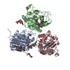
Type: D-saccharide, beta linking / Mass: 221.208 Da / Num. of mol.: 2 / Source method: isolated from a natural source / Formula: C8H15NO6
IdentifierTypeProgram
DGlcpNAcbCONDENSED IUPAC CARBOHYDRATE SYMBOLGMML 1.0
N-acetyl-b-D-glucopyranosamineCOMMON NAMEGMML 1.0
b-D-GlcpNAcIUPAC CARBOHYDRATE SYMBOLPDB-CARE 1.0
GlcNAcSNFG CARBOHYDRATE SYMBOLGMML 1.0

-
Non-polymers , 8 types, 676 molecules

#4: Chemical ChemComp-WSN / N-[(2R)-4-amino-1-imino-4-oxobutan-2-yl]-1-(1-{4-[(2,4-difluorophenyl)methoxy]phenyl}cyclopropane-1-carbonyl)-L-prolinamide


Mass: 498.522 Da / Num. of mol.: 3 / Source method: obtained synthetically / Formula: C26H28F2N4O4
#5: Chemical
ChemComp-EDO / 1,2-ETHANEDIOL / ETHYLENE GLYCOL


Mass: 62.068 Da / Num. of mol.: 4 / Source method: obtained synthetically / Formula: C2H6O2
#6: Chemical ChemComp-PG4 / TETRAETHYLENE GLYCOL


Mass: 194.226 Da / Num. of mol.: 1 / Source method: obtained synthetically / Formula: C8H18O5 / Comment: precipitant*YM
#7: Chemical ChemComp-EPE / 4-(2-HYDROXYETHYL)-1-PIPERAZINE ETHANESULFONIC ACID / HEPES


Mass: 238.305 Da / Num. of mol.: 1 / Source method: obtained synthetically / Formula: C8H18N2O4S / Comment: pH buffer*YM
#8: Chemical ChemComp-FLC / CITRATE ANION


Mass: 189.100 Da / Num. of mol.: 1 / Source method: obtained synthetically / Formula: C6H5O7
#9: Chemical ChemComp-MG / MAGNESIUM ION


Mass: 24.305 Da / Num. of mol.: 1 / Source method: obtained synthetically / Formula: Mg
#10: Chemical ChemComp-SO4 / SULFATE ION


Mass: 96.063 Da / Num. of mol.: 1 / Source method: obtained synthetically / Formula: SO4
#11: Water ChemComp-HOH / water


Mass: 18.015 Da / Num. of mol.: 664 / Source method: isolated from a natural source / Formula: H2O

-
Details

Has ligand of interestY

-
Experimental details

-
Experiment

ExperimentMethod: X-RAY DIFFRACTION / Number of used crystals: 1

-
Sample preparation

CrystalDensity Matthews: 2.01 Å3/Da / Density % sol: 38.88 %
Crystal growTemperature: 293 K / Method: vapor diffusion, sitting drop / pH: 7.8
Details: 25.7mg/mL protein in 25mM HEPES/NaOH pH7, 300 mM NaCl, 200mM Trehalose incubated with 10-fold excess of ligand, then mixed 50-70% with 50-30% reservoir consisting of 20% v/v PEG smear high, ...Details: 25.7mg/mL protein in 25mM HEPES/NaOH pH7, 300 mM NaCl, 200mM Trehalose incubated with 10-fold excess of ligand, then mixed 50-70% with 50-30% reservoir consisting of 20% v/v PEG smear high, 0.15M Li2SO4, 0.05M MgCL2, 0.1M HEPES/NaOH pH 7.8, total volume 200nL

-
Data collection

DiffractionMean temperature: 100 K
Diffraction sourceSource: SYNCHROTRON / Site: SLS / Beamline: X10SA / Wavelength: 0.99982 Å
DetectorType: MARMOSAIC 225 mm CCD / Detector: CCD / Date: Mar 23, 2021
RadiationProtocol: SINGLE WAVELENGTH / Monochromatic (M) / Laue (L): M / Scattering type: x-ray
Radiation wavelengthWavelength: 0.99982 Å / Relative weight: 1
ReflectionResolution: 1.7→112.62 Å / Num. obs: 128574 / % possible obs: 99 % / Redundancy: 3.02 % / Biso Wilson estimate: 35.81 Å2 / Rmerge(I) obs: 0.076 / Net I/σ(I): 8.32
Reflection shellResolution: 1.7→1.74 Å / Redundancy: 3 % / Rmerge(I) obs: 2.079 / Mean I/σ(I) obs: 0.49 / % possible all: 98.7

-
Processing

Software
NameVersionClassificationNB
REFMAC5.8.0267refinement
XSCALEdata scaling
PDB_EXTRACT3.27data extraction
XDSdata reduction
XSCALEdata scaling
PHASERphasing
RefinementMethod to determine structure: MOLECULAR REPLACEMENT
Starting model: INHOUSE MODEL

Resolution: 1.7→112.62 Å / Cor.coef. Fo:Fc: 0.96 / Cor.coef. Fo:Fc free: 0.944 / SU B: 2.341 / SU ML: 0.072 / Cross valid method: THROUGHOUT / σ(F): 0 / ESU R: 0.119 / ESU R Free: 0.114 / Stereochemistry target values: MAXIMUM LIKELIHOOD / Details: WITH A MG2+-CITRATE COMPLEX BOUND
RfactorNum. reflection% reflectionSelection details
Rfree0.201 4617 5.1 %RANDOM
Rwork0.169 ---
obs0.171 86420 70.2 %-
Solvent computationIon probe radii: 0.8 Å / Shrinkage radii: 0.8 Å / VDW probe radii: 1.2 Å / Solvent model: MASK
Displacement parametersBiso mean: 20.92 Å2
Baniso -1Baniso -2Baniso -3
1--0.07 Å20 Å20.04 Å2
2---0.03 Å20 Å2
3---0.08 Å2
Refinement stepCycle: LAST / Resolution: 1.7→112.62 Å
ProteinNucleic acidLigandSolventTotal
Num. atoms6341 0 227 664 7232
Refine LS restraints
Refine-IDTypeDev idealDev ideal targetNumber
X-RAY DIFFRACTIONr_bond_refined_d0.0080.0136922
X-RAY DIFFRACTIONr_bond_other_d0.0010.0176189
X-RAY DIFFRACTIONr_angle_refined_deg1.4431.6679385
X-RAY DIFFRACTIONr_angle_other_deg1.3261.62414303
X-RAY DIFFRACTIONr_dihedral_angle_1_deg6.8295812
X-RAY DIFFRACTIONr_dihedral_angle_2_deg36.71923.379364
X-RAY DIFFRACTIONr_dihedral_angle_3_deg13.166151112
X-RAY DIFFRACTIONr_dihedral_angle_4_deg10.4491527
X-RAY DIFFRACTIONr_chiral_restr0.0730.2872
X-RAY DIFFRACTIONr_gen_planes_refined0.0070.027815
X-RAY DIFFRACTIONr_gen_planes_other0.0010.021604
X-RAY DIFFRACTIONr_nbd_refined
X-RAY DIFFRACTIONr_nbd_other
X-RAY DIFFRACTIONr_nbtor_refined
X-RAY DIFFRACTIONr_nbtor_other
X-RAY DIFFRACTIONr_xyhbond_nbd_refined
X-RAY DIFFRACTIONr_xyhbond_nbd_other
X-RAY DIFFRACTIONr_metal_ion_refined
X-RAY DIFFRACTIONr_metal_ion_other
X-RAY DIFFRACTIONr_symmetry_vdw_refined
X-RAY DIFFRACTIONr_symmetry_vdw_other
X-RAY DIFFRACTIONr_symmetry_hbond_refined
X-RAY DIFFRACTIONr_symmetry_hbond_other
X-RAY DIFFRACTIONr_symmetry_metal_ion_refined
X-RAY DIFFRACTIONr_symmetry_metal_ion_other
X-RAY DIFFRACTIONr_mcbond_it1.241.9563173
X-RAY DIFFRACTIONr_mcbond_other1.2241.9553172
X-RAY DIFFRACTIONr_mcangle_it1.8912.9223962
X-RAY DIFFRACTIONr_mcangle_other
X-RAY DIFFRACTIONr_scbond_it
X-RAY DIFFRACTIONr_scbond_other
X-RAY DIFFRACTIONr_scangle_it
X-RAY DIFFRACTIONr_scangle_other
X-RAY DIFFRACTIONr_long_range_B_refined
X-RAY DIFFRACTIONr_long_range_B_other
X-RAY DIFFRACTIONr_rigid_bond_restr
X-RAY DIFFRACTIONr_sphericity_free
X-RAY DIFFRACTIONr_sphericity_bonded
LS refinement shellResolution: 1.7→1.74 Å / Total num. of bins used: 20
RfactorNum. reflection% reflection
Rfree0.281 16 -
Rwork0.27 467 -
obs--5.07 %

+
About Yorodumi

-
News

-
Feb 9, 2022. New format data for meta-information of EMDB entries

New format data for meta-information of EMDB entries

  • Version 3 of the EMDB header file is now the official format.
  • The previous official version 1.9 will be removed from the archive.

Related info.:EMDB header

External links:wwPDB to switch to version 3 of the EMDB data model

-
Aug 12, 2020. Covid-19 info

Covid-19 info

URL: https://pdbjlvh1.pdbj.org/emnavi/covid19.php

New page: Covid-19 featured information page in EM Navigator.

Related info.:Covid-19 info / Mar 5, 2020. Novel coronavirus structure data

+
Mar 5, 2020. Novel coronavirus structure data

Novel coronavirus structure data

Related info.:Yorodumi Speices / Aug 12, 2020. Covid-19 info

External links:COVID-19 featured content - PDBj / Molecule of the Month (242):Coronavirus Proteases

+
Jan 31, 2019. EMDB accession codes are about to change! (news from PDBe EMDB page)

EMDB accession codes are about to change! (news from PDBe EMDB page)

  • The allocation of 4 digits for EMDB accession codes will soon come to an end. Whilst these codes will remain in use, new EMDB accession codes will include an additional digit and will expand incrementally as the available range of codes is exhausted. The current 4-digit format prefixed with “EMD-” (i.e. EMD-XXXX) will advance to a 5-digit format (i.e. EMD-XXXXX), and so on. It is currently estimated that the 4-digit codes will be depleted around Spring 2019, at which point the 5-digit format will come into force.
  • The EM Navigator/Yorodumi systems omit the EMD- prefix.

Related info.:Q: What is EMD? / ID/Accession-code notation in Yorodumi/EM Navigator

External links:EMDB Accession Codes are Changing Soon! / Contact to PDBj

+
Jul 12, 2017. Major update of PDB

Major update of PDB

  • wwPDB released updated PDB data conforming to the new PDBx/mmCIF dictionary.
  • This is a major update changing the version number from 4 to 5, and with Remediation, in which all the entries are updated.
  • In this update, many items about electron microscopy experimental information are reorganized (e.g. em_software).
  • Now, EM Navigator and Yorodumi are based on the updated data.

External links:wwPDB Remediation / Enriched Model Files Conforming to OneDep Data Standards Now Available in the PDB FTP Archive

-
Yorodumi

Thousand views of thousand structures

  • Yorodumi is a browser for structure data from EMDB, PDB, SASBDB, etc.
  • This page is also the successor to EM Navigator detail page, and also detail information page/front-end page for Omokage search.
  • The word "yorodu" (or yorozu) is an old Japanese word meaning "ten thousand". "mi" (miru) is to see.

Related info.:EMDB / PDB / SASBDB / Comparison of 3 databanks / Yorodumi Search / Aug 31, 2016. New EM Navigator & Yorodumi / Yorodumi Papers / Jmol/JSmol / Function and homology information / Changes in new EM Navigator and Yorodumi

Read more