 Movie
Movie Controller

[English] 日本語
 Yorodumi
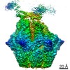
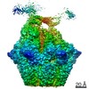
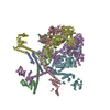
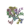
- PDB-7fiz: Processive cleavage of substrate at individual proteolytic active... -
+ Open data

- Basic information
| Entry | Database: PDB / ID: 7fiz | |||||||||||||||||||||
|---|---|---|---|---|---|---|---|---|---|---|---|---|---|---|---|---|---|---|---|---|---|---|
| Title | Processive cleavage of substrate at individual proteolytic active sites of the Lon protease complex (conformation 3) | |||||||||||||||||||||
 Components Components |
| |||||||||||||||||||||
 Keywords Keywords | HYDROLASE / AAA / protease / complex / proteolysis | |||||||||||||||||||||
| Function / homology |  Function and homology information Function and homology informationendopeptidase La / ATP-dependent peptidase activity / protein quality control for misfolded or incompletely synthesized proteins / cellular response to heat / sequence-specific DNA binding / serine-type endopeptidase activity / ATP hydrolysis activity / ATP binding / metal ion binding / identical protein binding / cytoplasm Similarity search - Function | |||||||||||||||||||||
| Biological species |  Meiothermus taiwanensis (bacteria)Meiothermus taiwanensis WR-220 (bacteria) Meiothermus taiwanensis (bacteria)Meiothermus taiwanensis WR-220 (bacteria) | |||||||||||||||||||||
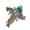
| Method | ELECTRON MICROSCOPY / single particle reconstruction / cryo EM / Resolution: 3.28 Å | |||||||||||||||||||||
 Authors Authors | Li, S. / Hsieh, K. / Kuo, C. / Su, S. / Huang, K. / Zhang, K. / Chang, C.I. | |||||||||||||||||||||
| Funding support |  Taiwan, 1items Taiwan, 1items
| |||||||||||||||||||||
 Citation Citation | Journal: Sci Adv / Year: 2021 Title: Processive cleavage of substrate at individual proteolytic active sites of the Lon protease complex. Authors: Shanshan Li / Kan-Yen Hsieh / Chiao-I Kuo / Shih-Chieh Su / Kai-Fa Huang / Kaiming Zhang / Chung-I Chang /  Abstract: The Lon protease is the prototype of a family of proteolytic machines with adenosine triphosphatase modules built into a substrate degradation chamber. Lon is known to degrade protein substrates in a ...The Lon protease is the prototype of a family of proteolytic machines with adenosine triphosphatase modules built into a substrate degradation chamber. Lon is known to degrade protein substrates in a processive fashion, cutting a protein chain processively into small peptides before commencing cleavages of another protein chain. Here, we present structural and biochemical evidence demonstrating that processive substrate degradation occurs at each of the six proteolytic active sites of Lon, which forms a deep groove that partially encloses the substrate polypeptide chain by accommodating only the unprimed residues and permits processive cleavage in the C-to-N direction. We identify a universally conserved acidic residue at the exit side of the binding groove indispensable for the proteolytic activity. This noncatalytic residue likely promotes processive proteolysis by carboxyl-carboxylate interactions with cleaved intermediates. Together, these results uncover a previously unrecognized mechanism for processive substrate degradation by the Lon protease. | |||||||||||||||||||||
| History |
|
- Structure visualization
| Movie |
Movie viewer |
|---|---|
| Structure viewer | Molecule: MolmilJmol/JSmol |
- Downloads & links
-Download
| PDBx/mmCIF format | 7fiz.cif.gz | 785.8 KB | Display | PDBx/mmCIF format |
|---|---|---|---|---|
| PDB format | pdb7fiz.ent.gz | 663.5 KB | Display | PDB format |
| PDBx/mmJSON format | 7fiz.json.gz | Tree view | PDBx/mmJSON format | |
| Others |  Other downloads Other downloads |
-Validation report
| Arichive directory | https://data.pdbj.org/pub/pdb/validation_reports/fi/7fizftp://data.pdbj.org/pub/pdb/validation_reports/fi/7fiz | HTTPS FTP |
|---|
-Related structure data
| Related structure data | 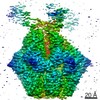 31607MC  7euxC  7euyC  7ev4C  7ev6C 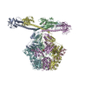 7fidC 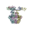 7fieC M: map data used to model this data C: citing same article ( |
|---|---|
| Similar structure data |


-Links
 PDBj
PDBj

- Assembly
| Deposited unit | 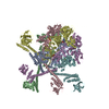
|
|---|---|
| 1 |
|
-Components
| #1: Protein | Mass: 90081.336 Da / Num. of mol.: 6 Source method: isolated from a genetically manipulated source Source: (gene. exp.) Meiothermus taiwanensis (bacteria) / Gene: lonA1, lon / Production host: #2: Protein/peptide | | Mass: 1890.321 Da / Num. of mol.: 1 Source method: isolated from a genetically manipulated source Source: (gene. exp.) Meiothermus taiwanensis WR-220 (bacteria)Production host: #3: Chemical | ChemComp-AGS /   Mass: 523.247 Da / Num. of mol.: 4 / Source method: obtained synthetically / Formula: C10H16N5O12P3S / Feature type: SUBJECT OF INVESTIGATION / Comment: ATP-gamma-S, energy-carrying molecule analogue*YM Mass: 523.247 Da / Num. of mol.: 4 / Source method: obtained synthetically / Formula: C10H16N5O12P3S / Feature type: SUBJECT OF INVESTIGATION / Comment: ATP-gamma-S, energy-carrying molecule analogue*YM#4: Chemical | ChemComp-ADP / |   Mass: 427.201 Da / Num. of mol.: 1 / Source method: obtained synthetically / Formula: C10H15N5O10P2 / Feature type: SUBJECT OF INVESTIGATION / Comment: ADP, energy-carrying molecule*YM Mass: 427.201 Da / Num. of mol.: 1 / Source method: obtained synthetically / Formula: C10H15N5O10P2 / Feature type: SUBJECT OF INVESTIGATION / Comment: ADP, energy-carrying molecule*YMHas ligand of interest | Y | Has protein modification | N | |
|---|
-Experimental details
-Experiment
| Experiment | Method: ELECTRON MICROSCOPY |
|---|---|
| EM experiment | Aggregation state: PARTICLE / 3D reconstruction method: single particle reconstruction |
- Sample preparation
| Component | Name: Lon bound to endogenous substrate / Type: COMPLEX / Entity ID: #1-#2 / Source: RECOMBINANT |
|---|---|
| Molecular weight | Value: 0.6 MDa / Experimental value: YES |
| Source (natural) | Organism: Meiothermus taiwanensis WR-220 (bacteria) |
| Source (recombinant) | Organism: |
| Buffer solution | pH: 7.5 |
| Specimen | Embedding applied: NO / Shadowing applied: NO / Staining applied: NO / Vitrification applied: YES |
| Specimen support | Grid material: COPPER / Grid mesh size: 200 divisions/in. / Grid type: Quantifoil R1.2/1.3 |
| Vitrification | Cryogen name: ETHANE |
- Electron microscopy imaging
| Experimental equipment |  Model: Titan Krios / Image courtesy: FEI Company |
|---|---|
| Microscopy | Model: FEI TITAN KRIOS |
| Electron gun | Electron source:  FIELD EMISSION GUN / Accelerating voltage: 300 kV / Illumination mode: FLOOD BEAM FIELD EMISSION GUN / Accelerating voltage: 300 kV / Illumination mode: FLOOD BEAM |
| Electron lens | Mode: BRIGHT FIELD |
| Image recording | Electron dose: 48 e/Å2 / Film or detector model: FEI FALCON IV (4k x 4k) |
- Processing
| Software | Name: PHENIX / Version: 1.13_2998: / Classification: refinement | ||||||||||||||||||||||||
|---|---|---|---|---|---|---|---|---|---|---|---|---|---|---|---|---|---|---|---|---|---|---|---|---|---|
| EM software |
| ||||||||||||||||||||||||
| CTF correction | Type: NONE | ||||||||||||||||||||||||
| Symmetry | Point symmetry: C1 (asymmetric) | ||||||||||||||||||||||||
| 3D reconstruction | Resolution: 3.28 Å / Resolution method: FSC 0.143 CUT-OFF / Num. of particles: 77159 / Symmetry type: POINT | ||||||||||||||||||||||||
| Refine LS restraints |
|
