 Movie
Movie Controller

[English] 日本語
 Yorodumi
- PDB-7fd5: A complete three-dimensional structure of the Lon protease transl... -
+ Open data

- Basic information
| Entry | Database: PDB / ID: 7fd5 | ||||||
|---|---|---|---|---|---|---|---|
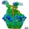
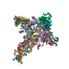
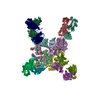
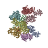
| Title | A complete three-dimensional structure of the Lon protease translocating a protein substrate (conformation 2) | ||||||
 Components Components |
| ||||||
 Keywords Keywords | HYDROLASE/PROTEIN BINDING / AAA+ protease / Lon / complete three-dimensional structure / N-terminal domain / CYTOSOLIC PROTEIN / HYDROLASE-PROTEIN BINDING complex | ||||||
| Function / homology |  Function and homology information Function and homology informationendopeptidase La / ATP-dependent peptidase activity / protein quality control for misfolded or incompletely synthesized proteins / cellular response to heat / sequence-specific DNA binding / serine-type endopeptidase activity / ATP hydrolysis activity / ATP binding / metal ion binding / identical protein binding / cytoplasm Similarity search - Function | ||||||
| Biological species |  Meiothermus taiwanensis (bacteria) Meiothermus taiwanensis (bacteria) | ||||||
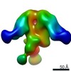
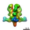
| Method | ELECTRON MICROSCOPY / single particle reconstruction / cryo EM / Resolution: 2.4 Å | ||||||
 Authors Authors | Li, S. / Hsieh, K. / Kuo, C. / Lee, S. / Pintilie, G. / Zhang, K. / Chang, C. | ||||||
| Funding support |  Taiwan, 1items Taiwan, 1items
| ||||||
 Citation Citation | Journal: Sci Adv / Year: 2021 Title: Complete three-dimensional structures of the Lon protease translocating a protein substrate. Authors: Shanshan Li / Kan-Yen Hsieh / Chiao-I Kuo / Szu-Hui Lee / Grigore D Pintilie / Kaiming Zhang / Chung-I Chang /   Abstract: Lon is an evolutionarily conserved proteolytic machine carrying out a wide spectrum of biological activities by degrading misfolded damaged proteins and specific cellular substrates. Lon contains a ...Lon is an evolutionarily conserved proteolytic machine carrying out a wide spectrum of biological activities by degrading misfolded damaged proteins and specific cellular substrates. Lon contains a large N-terminal domain and forms a hexameric core of fused adenosine triphosphatase and protease domains. Here, we report two complete structures of Lon engaging a substrate, determined by cryo–electron microscopy to 2.4-angstrom resolution. These structures show a multilayered architecture featuring a tensegrity triangle complex, uniquely constructed by six long N-terminal helices. The interlocked helix triangle is assembled on the top of the hexameric core to spread a web of six globular substrate-binding domains. It serves as a multipurpose platform that controls the access of substrates to the AAA+ ring, provides a ruler-based mechanism for substrate selection, and acts as a pulley device to facilitate unfolding of the translocated substrate. This work provides a complete framework for understanding the structural mechanisms of Lon. | ||||||
| History |
|
- Structure visualization
| Movie |
Movie viewer |
|---|---|
| Structure viewer | Molecule: MolmilJmol/JSmol |
- Downloads & links
-Download
| PDBx/mmCIF format | 7fd5.cif.gz | 787.6 KB | Display | PDBx/mmCIF format |
|---|---|---|---|---|
| PDB format | pdb7fd5.ent.gz | 665.5 KB | Display | PDB format |
| PDBx/mmJSON format | 7fd5.json.gz | Tree view | PDBx/mmJSON format | |
| Others |  Other downloads Other downloads |
-Validation report
| Arichive directory | https://data.pdbj.org/pub/pdb/validation_reports/fd/7fd5ftp://data.pdbj.org/pub/pdb/validation_reports/fd/7fd5 | HTTPS FTP |
|---|
-Related structure data
| Related structure data | 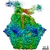 31535MC 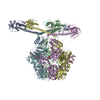 7fd4C M: map data used to model this data C: citing same article ( |
|---|---|
| Similar structure data |

-Links
 PDBj
PDBj


- Assembly
| Deposited unit | 
|
|---|---|
| 1 |
|
-Components
| #1: Protein | Mass: 88554.594 Da / Num. of mol.: 6 Source method: isolated from a genetically manipulated source Source: (gene. exp.) Meiothermus taiwanensis (bacteria) / Gene: lonA1, lon / Production host: #2: Protein/peptide | | Mass: 1890.321 Da / Num. of mol.: 1 / Source method: isolated from a natural source / Source: (natural) #3: Chemical |   Mass: 427.201 Da / Num. of mol.: 3 / Source method: obtained synthetically / Formula: C10H15N5O10P2 / Feature type: SUBJECT OF INVESTIGATION / Comment: ADP, energy-carrying molecule*YM Mass: 427.201 Da / Num. of mol.: 3 / Source method: obtained synthetically / Formula: C10H15N5O10P2 / Feature type: SUBJECT OF INVESTIGATION / Comment: ADP, energy-carrying molecule*YM#4: Chemical | ChemComp-4KZ /   Mass: 418.253 Da / Num. of mol.: 6 / Source method: obtained synthetically / Formula: C22H23BN4O4 / Feature type: SUBJECT OF INVESTIGATION Mass: 418.253 Da / Num. of mol.: 6 / Source method: obtained synthetically / Formula: C22H23BN4O4 / Feature type: SUBJECT OF INVESTIGATION#5: Chemical |   Mass: 523.247 Da / Num. of mol.: 3 / Source method: obtained synthetically / Formula: C10H16N5O12P3S / Feature type: SUBJECT OF INVESTIGATION / Comment: ATP-gamma-S, energy-carrying molecule analogue*YM Mass: 523.247 Da / Num. of mol.: 3 / Source method: obtained synthetically / Formula: C10H16N5O12P3S / Feature type: SUBJECT OF INVESTIGATION / Comment: ATP-gamma-S, energy-carrying molecule analogue*YMHas ligand of interest | Y | Has protein modification | Y | Sequence details | Authors know the sequence of chain S (UNP P02662) but don't know how the coordinates align with the ...Authors know the sequence of chain S (UNP P02662) but don't know how the coordinates align with the sequence. P02662 sequence is MKLLILTCLV | |
|---|
-Experimental details
-Experiment
| Experiment | Method: ELECTRON MICROSCOPY |
|---|---|
| EM experiment | Aggregation state: PARTICLE / 3D reconstruction method: single particle reconstruction |
- Sample preparation
| Component |
| ||||||||||||||||||||||||
|---|---|---|---|---|---|---|---|---|---|---|---|---|---|---|---|---|---|---|---|---|---|---|---|---|---|
| Molecular weight | Value: 0.37 MDa / Experimental value: YES | ||||||||||||||||||||||||
| Source (natural) |
| ||||||||||||||||||||||||
| Source (recombinant) | Organism: | ||||||||||||||||||||||||
| Buffer solution | pH: 8 | ||||||||||||||||||||||||
| Specimen | Conc.: 0.5 mg/ml / Embedding applied: NO / Shadowing applied: NO / Staining applied: NO / Vitrification applied: YES | ||||||||||||||||||||||||
| Specimen support | Grid material: COPPER / Grid type: Quantifoil R2/1 | ||||||||||||||||||||||||
| Vitrification | Cryogen name: ETHANE |
- Electron microscopy imaging
| Experimental equipment |  Model: Titan Krios / Image courtesy: FEI Company |
|---|---|
| Microscopy | Model: FEI TITAN KRIOS |
| Electron gun | Electron source:  FIELD EMISSION GUN / Accelerating voltage: 300 kV / Illumination mode: FLOOD BEAM FIELD EMISSION GUN / Accelerating voltage: 300 kV / Illumination mode: FLOOD BEAM |
| Electron lens | Mode: BRIGHT FIELD |
| Image recording | Electron dose: 48 e/Å2 / Film or detector model: FEI FALCON IV (4k x 4k) |
- Processing
| CTF correction | Type: NONE |
|---|---|
| 3D reconstruction | Resolution: 2.4 Å / Resolution method: FSC 0.143 CUT-OFF / Num. of particles: 156330 / Symmetry type: POINT |
