 Movie
Movie Controller


+ Open data

- Basic information
| Entry | Database: PDB / ID: 7ffh | ||||||
|---|---|---|---|---|---|---|---|
| Title | Diarylpentanoid-producing polyketide synthase (N199L mutant) | ||||||
 Components Components | Type III polyketide synthase | ||||||
 Keywords Keywords | TRANSFERASE / diarylpentanoid / type-III polyketide synthase | ||||||
| Function / homology |  Function and homology information Function and homology informationpolyketide biosynthetic process / acyltransferase activity, transferring groups other than amino-acyl groups / endoplasmic reticulum Similarity search - Function | ||||||
| Biological species |  Aquilaria sinensis (plant) Aquilaria sinensis (plant) | ||||||
| Method |  X-RAY DIFFRACTION / SYNCHROTRON / MOLECULAR REPLACEMENT / Resolution: 2.2 Å X-RAY DIFFRACTION / SYNCHROTRON / MOLECULAR REPLACEMENT / Resolution: 2.2 Å | ||||||
 Authors Authors | Morita, H. / Wong, C.P. / Liu, Q. / Takeshi, K. / Lee, Y. / Nakashima, Y. | ||||||
 Citation Citation | Journal: Nat Commun / Year: 2022 Title: Identification of a diarylpentanoid-producing polyketide synthase revealing an unusual biosynthetic pathway of 2-(2-phenylethyl)chromones in agarwood. Authors: Wang, X.H. / Gao, B.W. / Nakashima, Y. / Mori, T. / Zhang, Z.X. / Kodama, T. / Lee, Y.E. / Zhang, Z.K. / Wong, C.P. / Liu, Q.Q. / Qi, B.W. / Wang, J. / Li, J. / Liu, X. / Abe, I. / Morita, H. ...Authors: Wang, X.H. / Gao, B.W. / Nakashima, Y. / Mori, T. / Zhang, Z.X. / Kodama, T. / Lee, Y.E. / Zhang, Z.K. / Wong, C.P. / Liu, Q.Q. / Qi, B.W. / Wang, J. / Li, J. / Liu, X. / Abe, I. / Morita, H. / Tu, P.F. / Shi, S.P. | ||||||
| History |
|
- Structure visualization
| Structure viewer | Molecule: MolmilJmol/JSmol |
|---|
- Downloads & links
-Download
| PDBx/mmCIF format | 7ffh.cif.gz | 164 KB | Display | PDBx/mmCIF format |
|---|---|---|---|---|
| PDB format | pdb7ffh.ent.gz | 124.5 KB | Display | PDB format |
| PDBx/mmJSON format | 7ffh.json.gz | Tree view | PDBx/mmJSON format | |
| Others |  Other downloads Other downloads |
-Validation report
| Arichive directory | https://data.pdbj.org/pub/pdb/validation_reports/ff/7ffhftp://data.pdbj.org/pub/pdb/validation_reports/ff/7ffh | HTTPS FTP |
|---|
-Related structure data
| Related structure data |  7ffaC  7ffcC  7ffgC  7ffiC  4yjyS S: Starting model for refinement C: citing same article ( |
|---|---|
| Similar structure data |


-Links
 PDBj
PDBj

- Assembly
| Deposited unit | 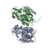
| ||||||||||||
|---|---|---|---|---|---|---|---|---|---|---|---|---|---|
| 1 | 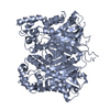
| ||||||||||||
| 2 | 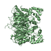
| ||||||||||||
| Unit cell |
| ||||||||||||
| Components on special symmetry positions |
|
-Components
| #1: Protein | Mass: 46844.656 Da / Num. of mol.: 2 / Mutation: N199L Source method: isolated from a genetically manipulated source Source: (gene. exp.) Aquilaria sinensis (plant) / Production host:  #2: Water | ChemComp-HOH / |  Mass: 18.015 Da / Num. of mol.: 88 / Source method: isolated from a natural source / Formula: H2O Mass: 18.015 Da / Num. of mol.: 88 / Source method: isolated from a natural source / Formula: H2O |
|---|
-Experimental details
-Experiment
| Experiment | Method: X-RAY DIFFRACTION / Number of used crystals: 1 |
|---|
- Sample preparation
| Crystal | Density Matthews: 2.17 Å3/Da / Density % sol: 43.38 % |
|---|---|
| Crystal grow | Temperature: 293 K / Method: vapor diffusion, sitting drop Details: 0.1M Tris-HCl, pH 8.5, 25% PEG 8000, 0.3M KF, 4% butanediol |
-Data collection
| Diffraction | Mean temperature: 100 K / Serial crystal experiment: N |
|---|---|
| Diffraction source | Source: SYNCHROTRON / Site: Photon Factory  / Beamline: BL-5A / Wavelength: 1 Å / Beamline: BL-5A / Wavelength: 1 Å |
| Detector | Type: DECTRIS PILATUS3 S 6M / Detector: PIXEL / Date: May 18, 2019 |
| Radiation | Protocol: SINGLE WAVELENGTH / Monochromatic (M) / Laue (L): M / Scattering type: x-ray |
| Radiation wavelength | Wavelength: 1 Å / Relative weight: 1 |
| Reflection | Resolution: 2.2→42.64 Å / Num. obs: 40917 / % possible obs: 99.7 % / Redundancy: 4.4 % / Biso Wilson estimate: 42.58 Å2 / CC1/2: 0.999 / Rmerge(I) obs: 0.043 / Net I/σ(I): 15.1 |
| Reflection shell | Resolution: 2.2→2.27 Å / Redundancy: 4.5 % / Rmerge(I) obs: 0.36 / Mean I/σ(I) obs: 3.3 / Num. unique obs: 3522 / CC1/2: 0.954 / % possible all: 99.7 |
- Processing
| Software |
| ||||||||||||||||||||||||||||||||||||||||||||||||||||||||||||||||||||||||||||||||||||||||||||||||||||||||||||||||
|---|---|---|---|---|---|---|---|---|---|---|---|---|---|---|---|---|---|---|---|---|---|---|---|---|---|---|---|---|---|---|---|---|---|---|---|---|---|---|---|---|---|---|---|---|---|---|---|---|---|---|---|---|---|---|---|---|---|---|---|---|---|---|---|---|---|---|---|---|---|---|---|---|---|---|---|---|---|---|---|---|---|---|---|---|---|---|---|---|---|---|---|---|---|---|---|---|---|---|---|---|---|---|---|---|---|---|---|---|---|---|---|---|---|
| Refinement | Method to determine structure: MOLECULAR REPLACEMENT Starting model: 4YJY Resolution: 2.2→40.51 Å / SU ML: 0.2724 / Cross valid method: FREE R-VALUE / σ(F): 1.34 / Phase error: 31.5323 Stereochemistry target values: GeoStd + Monomer Library + CDL v1.2
| ||||||||||||||||||||||||||||||||||||||||||||||||||||||||||||||||||||||||||||||||||||||||||||||||||||||||||||||||
| Solvent computation | Shrinkage radii: 0.9 Å / VDW probe radii: 1.11 Å / Solvent model: FLAT BULK SOLVENT MODEL | ||||||||||||||||||||||||||||||||||||||||||||||||||||||||||||||||||||||||||||||||||||||||||||||||||||||||||||||||
| Displacement parameters | Biso mean: 61.81 Å2 | ||||||||||||||||||||||||||||||||||||||||||||||||||||||||||||||||||||||||||||||||||||||||||||||||||||||||||||||||
| Refinement step | Cycle: LAST / Resolution: 2.2→40.51 Å
| ||||||||||||||||||||||||||||||||||||||||||||||||||||||||||||||||||||||||||||||||||||||||||||||||||||||||||||||||
| Refine LS restraints |
| ||||||||||||||||||||||||||||||||||||||||||||||||||||||||||||||||||||||||||||||||||||||||||||||||||||||||||||||||
| LS refinement shell |
|
