 Movie
Movie Controller


+ Open data

- Basic information
| Entry | Database: PDB / ID: 7f37 | |||||||||||||||
|---|---|---|---|---|---|---|---|---|---|---|---|---|---|---|---|---|
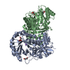
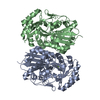
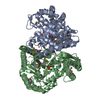
| Title | Crystal structure of AtaT2-AtaR2 complex | |||||||||||||||
 Components Components |
| |||||||||||||||
 Keywords Keywords | TRANSFERASE / Acetyltransferase / TOXIN | |||||||||||||||
| Function / homology | : / :  Function and homology information Function and homology information | |||||||||||||||
| Biological species |  | |||||||||||||||
| Method |  X-RAY DIFFRACTION / SYNCHROTRON / MOLECULAR REPLACEMENT / Resolution: 2.896 Å X-RAY DIFFRACTION / SYNCHROTRON / MOLECULAR REPLACEMENT / Resolution: 2.896 Å | |||||||||||||||
 Authors Authors | Yashiro, Y. / Tomita, K. | |||||||||||||||
| Funding support |  Japan, 4items Japan, 4items
| |||||||||||||||
 Citation Citation | Journal: Cell Rep / Year: 2021 Title: Molecular basis of glycyl-tRNAGly acetylation by TacT from Salmonella Typhimurium Authors: Yashiro, Y. / Zhang, C. / Sakaguchi, Y. / Suzuki, T. / Tomita, K. | |||||||||||||||
| History |
|
- Structure visualization
| Structure viewer | Molecule: MolmilJmol/JSmol |
|---|
- Downloads & links
-Download
| PDBx/mmCIF format | 7f37.cif.gz | 138.5 KB | Display | PDBx/mmCIF format |
|---|---|---|---|---|
| PDB format | pdb7f37.ent.gz | 108.1 KB | Display | PDB format |
| PDBx/mmJSON format | 7f37.json.gz | Tree view | PDBx/mmJSON format | |
| Others |  Other downloads Other downloads |
-Validation report
| Summary document | 7f37_validation.pdf.gz | 478.7 KB | Display | wwPDB validaton report |
|---|---|---|---|---|
| Full document | 7f37_full_validation.pdf.gz | 501.3 KB | Display | |
| Data in XML | 7f37_validation.xml.gz | 26.2 KB | Display | |
| Data in CIF | 7f37_validation.cif.gz | 35.4 KB | Display | |
| Arichive directory | https://data.pdbj.org/pub/pdb/validation_reports/f3/7f37ftp://data.pdbj.org/pub/pdb/validation_reports/f3/7f37 | HTTPS FTP |
-Related structure data
| Related structure data |  7f36C 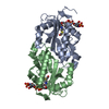 5fvjS S: Starting model for refinement C: citing same article ( |
|---|---|
| Similar structure data |


-Links
 PDBj
PDBj- Assembly
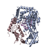
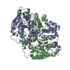
| Deposited unit | 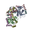
| ||||||||
|---|---|---|---|---|---|---|---|---|---|
| 1 |
| ||||||||
| Unit cell |
|
-Components
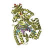
| #1: Protein | Mass: 18741.707 Da / Num. of mol.: 2 Source method: isolated from a genetically manipulated source Source: (gene. exp.) Gene: Z4777, CQJ22_000873, E3157_02175, E3158_02185, E5F07_24590, FDZ86_02175 Production host: #2: Protein | Mass: 10433.930 Da / Num. of mol.: 4 Source method: isolated from a genetically manipulated source Source: (gene. exp.) Gene: CQJ22_000874, E3157_02180, E3158_02190, E3175_02180, E5F07_24585, FDZ86_02180 Production host: #3: Water | ChemComp-HOH / |  Mass: 18.015 Da / Num. of mol.: 6 / Source method: isolated from a natural source / Formula: H2O Mass: 18.015 Da / Num. of mol.: 6 / Source method: isolated from a natural source / Formula: H2O |
|---|
-Experimental details
-Experiment
| Experiment | Method: X-RAY DIFFRACTION / Number of used crystals: 1 |
|---|
- Sample preparation
| Crystal | Density Matthews: 2.5 Å3/Da / Density % sol: 50.85 % |
|---|---|
| Crystal grow | Temperature: 293 K / Method: vapor diffusion, sitting drop / Details: 0.2M Potassium formate, 20% PEG 3350 |
-Data collection
| Diffraction | Mean temperature: 100 K / Serial crystal experiment: N |
|---|---|
| Diffraction source | Source: SYNCHROTRON / Site: Photon Factory / Beamline: BL-17A / Wavelength: 0.98 Å |
| Detector | Type: DECTRIS EIGER X 16M / Detector: PIXEL / Date: Nov 2, 2020 |
| Radiation | Protocol: SINGLE WAVELENGTH / Monochromatic (M) / Laue (L): M / Scattering type: x-ray |
| Radiation wavelength | Wavelength: 0.98 Å / Relative weight: 1 |
| Reflection | Resolution: 2.896→46.12 Å / Num. obs: 17558 / % possible obs: 99.9 % / Redundancy: 13.4 % / CC1/2: 0.993 / Rmerge(I) obs: 0.28 / Net I/σ(I): 7.5 |
| Reflection shell | Resolution: 2.9→3 Å / Rmerge(I) obs: 1.92 / Mean I/σ(I) obs: 1.4 / Num. unique obs: 1708 / CC1/2: 0.76 |
- Processing
| Software |
| ||||||||||||||||||||||||||||||||||||
|---|---|---|---|---|---|---|---|---|---|---|---|---|---|---|---|---|---|---|---|---|---|---|---|---|---|---|---|---|---|---|---|---|---|---|---|---|---|
| Refinement | Method to determine structure: MOLECULAR REPLACEMENT Starting model: 5fvj Resolution: 2.896→45.375 Å / SU ML: 0.41 / Cross valid method: THROUGHOUT / σ(F): 1.36 / Phase error: 30.3 / Stereochemistry target values: ML Details: The reflection data set was anisotropically truncated and corrected using UCLA-DOE LAB Diffraction Anisotropy Server, and used for the refinement. Also, PDB extract was run using the ...Details: The reflection data set was anisotropically truncated and corrected using UCLA-DOE LAB Diffraction Anisotropy Server, and used for the refinement. Also, PDB extract was run using the anisotropically processed structure factor (.mtz) file.
| ||||||||||||||||||||||||||||||||||||
| Solvent computation | Shrinkage radii: 0.9 Å / VDW probe radii: 1.11 Å / Solvent model: FLAT BULK SOLVENT MODEL | ||||||||||||||||||||||||||||||||||||
| Displacement parameters | Biso max: 105.73 Å2 / Biso mean: 37.6895 Å2 / Biso min: 3.85 Å2 | ||||||||||||||||||||||||||||||||||||
| Refinement step | Cycle: final / Resolution: 2.896→45.375 Å
| ||||||||||||||||||||||||||||||||||||
| LS refinement shell | Refine-ID: X-RAY DIFFRACTION / Rfactor Rfree error: 0
|
