- PDB-5fvj: Crystal structure of TacT (tRNA acetylating toxin) from Salmonella -
+
Open data
ID or keywords:
Loading...
-
Basic information
Entry
Database: PDB / ID: 5fvj
Title
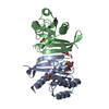
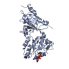
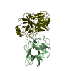
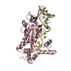
Crystal structure of TacT (tRNA acetylating toxin) from Salmonella
Components
PUTATIVE ACETYLTRANSFERASE
Keywords
TRANSFERASE / ACETYLTRANSFERASE
Function / homology
Function and homology information
acyltransferase activity, transferring groups other than amino-acyl groups / Transferases; Acyltransferases; Transferring groups other than aminoacyl groups / tRNA binding / DNA-templated transcription Similarity search - Function
Protocol: SINGLE WAVELENGTH / Monochromatic (M) / Laue (L): M / Scattering type: x-ray
Radiation wavelength
Wavelength: 0.92819 Å / Relative weight: 1
Reflection
Resolution: 1.7→55.2 Å / Num. obs: 32830 / % possible obs: 96.2 % / Observed criterion σ(I): -4 / Redundancy: 6.2 % / Rmerge(I) obs: 0.14 / Net I/σ(I): 8.6
Reflection shell
Resolution: 1.7→1.79 Å / Redundancy: 6 % / Rmerge(I) obs: 0.84 / Mean I/σ(I) obs: 2 / % possible all: 96.5
-
Processing
Software
Name
Version
Classification
REFMAC
5.8.0107
refinement
MOSFLM
datareduction
SCALA
datascaling
PHENIX
phasing
Refinement
Method to determine structure: SAD Starting model: NONE Resolution: 1.7→55.23 Å / Cor.coef. Fo:Fc: 0.961 / Cor.coef. Fo:Fc free: 0.934 / SU B: 7.044 / SU ML: 0.1 / Cross valid method: THROUGHOUT / ESU R: 0.295 / ESU R Free: 0.125 / Stereochemistry target values: MAXIMUM LIKELIHOOD Details: HYDROGENS HAVE BEEN ADDED IN THE RIDING POSITIONS. U VALUES REFINED INDIVIDUALLY
Rfactor
Num. reflection
% reflection
Selection details
Rfree
0.2254
1676
5.1 %
RANDOM
Rwork
0.17164
-
-
-
obs
0.17447
30992
95.53 %
-
Solvent computation
Ion probe radii: 0.8 Å / Shrinkage radii: 0.8 Å / VDW probe radii: 1.2 Å / Solvent model: MASK
 Movie
Movie Controller

 Yorodumi
 Open data
 Basic information
 Components
 Keywords
 Function and homology information
 SALMONELLA ENTERICA SUBSP. ENTERICA SEROVAR TYPHIMURIUM (bacteria)
 X-RAY DIFFRACTION /
  Authors
 Citation
 Structure visualization
 Downloads & links
 Other downloads


 PDBj
PDBj

 Assembly

 Mass: 809.571 Da / Num. of mol.: 2 / Source method: obtained synthetically / Formula: C23H38N7O17P3S
 Mass: 18.015 Da / Num. of mol.: 426 / Source method: isolated from a natural source / Formula: H2O
 Sample preparation
 / Beamline: I04-1 / Wavelength: 0.92819
  Processing