 Movie
Movie Controller

[English] 日本語
 Yorodumi
- PDB-7f18: Crystal Structure of a mutant of acid phosphatase from Pseudomona... -
+ Open data

- Basic information
| Entry | Database: PDB / ID: 7f18 | ||||||
|---|---|---|---|---|---|---|---|
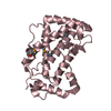
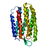
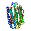
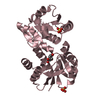
| Title | Crystal Structure of a mutant of acid phosphatase from Pseudomonas aeruginosa (Q57H/W58P/D135R) | ||||||
 Components Components | Acid phosphatase | ||||||
 Keywords Keywords | HYDROLASE / phosphorylation hydrolysis | ||||||
| Function / homology | Acid phosphatase, class A, bacterial / Acid phosphatase homologues / Phosphatidic acid phosphatase type 2/haloperoxidase / PAP2 superfamily / Phosphatidic acid phosphatase type 2/haloperoxidase superfamily / acid phosphatase / acid phosphatase activity / outer membrane-bounded periplasmic space / Acid phosphatase Function and homology information Function and homology information | ||||||
| Biological species |   Pseudomonas aeruginosa (bacteria) Pseudomonas aeruginosa (bacteria) | ||||||
| Method |  X-RAY DIFFRACTION / MOLECULAR REPLACEMENT / Resolution: 3.3 Å X-RAY DIFFRACTION / MOLECULAR REPLACEMENT / Resolution: 3.3 Å | ||||||
 Authors Authors | Xu, X. / Hou, X.D. / Song, W. / Yin, D.J. / Rao, Y.J. / Liu, L.M. | ||||||
| Funding support |  China, 1items China, 1items
| ||||||
 Citation Citation | Journal: Acs Catalysis / Year: 2021 Title: Local Electric Field Modulated Reactivity of Pseudomonas aeruginosa Acid Phosphatase for Enhancing Phosphorylation of l-Ascorbic Acid Authors: Xu, X. / Yan, S. / Hou, X. / Song, W. / Wang, L. / Wu, T. / Qi, M. / Wu, J. / Rao, Y. / Wang, B. / Liu, L. | ||||||
| History |
|
- Structure visualization
| Structure viewer | Molecule: MolmilJmol/JSmol |
|---|
- Downloads & links
-Download
| PDBx/mmCIF format | 7f18.cif.gz | 129.7 KB | Display | PDBx/mmCIF format |
|---|---|---|---|---|
| PDB format | pdb7f18.ent.gz | 102.5 KB | Display | PDB format |
| PDBx/mmJSON format | 7f18.json.gz | Tree view | PDBx/mmJSON format | |
| Others |  Other downloads Other downloads |
-Validation report
| Summary document | 7f18_validation.pdf.gz | 444.6 KB | Display | wwPDB validaton report |
|---|---|---|---|---|
| Full document | 7f18_full_validation.pdf.gz | 457.1 KB | Display | |
| Data in XML | 7f18_validation.xml.gz | 23.6 KB | Display | |
| Data in CIF | 7f18_validation.cif.gz | 31.7 KB | Display | |
| Arichive directory | https://data.pdbj.org/pub/pdb/validation_reports/f1/7f18ftp://data.pdbj.org/pub/pdb/validation_reports/f1/7f18 | HTTPS FTP |
-Related structure data
| Related structure data |  7f17SC S: Starting model for refinement C: citing same article ( |
|---|---|
| Similar structure data |
-Links
 PDBj
PDBj
- Assembly
| Deposited unit | 
| ||||||||
|---|---|---|---|---|---|---|---|---|---|
| 1 | 
| ||||||||
| 2 | 
| ||||||||
| 3 | 
| ||||||||
| Unit cell |
|
-Components
| #1: Protein | Mass: 23559.756 Da / Num. of mol.: 3 / Mutation: Q57H/W58P/D135R Source method: isolated from a genetically manipulated source Source: (gene. exp.) Pseudomonas aeruginosa (bacteria)Gene: phoC, C0044_01345, CAZ10_16160, DT376_10070, DZ962_30070, IPC116_25940, IPC1295_25655, IPC1323_25265, IPC1481_23795, IPC1505_26850, IPC1509_05755, IPC36_01875, IPC582_09790, IPC620_15205, ...Gene: phoC, C0044_01345, CAZ10_16160, DT376_10070, DZ962_30070, IPC116_25940, IPC1295_25655, IPC1323_25265, IPC1481_23795, IPC1505_26850, IPC1509_05755, IPC36_01875, IPC582_09790, IPC620_15205, IPC669_21580, NCTC13621_04764, RW109_RW109_00792 Production host: |
|---|
-Experimental details
-Experiment
| Experiment | Method: X-RAY DIFFRACTION / Number of used crystals: 1 |
|---|
- Sample preparation
| Crystal | Density Matthews: 2.81 Å3/Da / Density % sol: 56.17 % |
|---|---|
| Crystal grow | Temperature: 288.15 K / Method: vapor diffusion, sitting drop Details: 0.1M BIS-TRIS pH 6.5, 50% v/v Polypropylene glycol P 400 |
-Data collection
| Diffraction | Mean temperature: 100 K / Serial crystal experiment: N |
|---|---|
| Diffraction source | Source: SEALED TUBE / Type: BRUKER D8 QUEST / Wavelength: 1.542 Å |
| Detector | Type: Bruker PHOTON II / Detector: PIXEL / Date: Mar 2, 2021 |
| Radiation | Protocol: SINGLE WAVELENGTH / Monochromatic (M) / Laue (L): M / Scattering type: x-ray |
| Radiation wavelength | Wavelength: 1.542 Å / Relative weight: 1 |
| Reflection | Resolution: 3.189→23.63 Å / Num. obs: 13485 / % possible obs: 99.07 % / Redundancy: 4.92 % / Rsym value: 0.126 / Net I/av σ(I): 30.468 / Net I/σ(I): 9.9 |
| Reflection shell | Resolution: 3.26→3.52 Å / Num. unique obs: 855 / Rsym value: 0.394 |
- Processing
| Software |
| ||||||||||||||||||||||||||||||||||||||||||||||||||||||||||||
|---|---|---|---|---|---|---|---|---|---|---|---|---|---|---|---|---|---|---|---|---|---|---|---|---|---|---|---|---|---|---|---|---|---|---|---|---|---|---|---|---|---|---|---|---|---|---|---|---|---|---|---|---|---|---|---|---|---|---|---|---|---|
| Refinement | Method to determine structure: MOLECULAR REPLACEMENT Starting model: 7F17 Resolution: 3.3→23.63 Å / Cor.coef. Fo:Fc: 0.955 / Cor.coef. Fo:Fc free: 0.892 / SU B: 28.836 / SU ML: 0.437 / Cross valid method: THROUGHOUT / σ(F): 0 / ESU R Free: 0.6 / Stereochemistry target values: MAXIMUM LIKELIHOOD Details: HYDROGENS HAVE BEEN ADDED IN THE RIDING POSITIONS U VALUES : REFINED INDIVIDUALLY
| ||||||||||||||||||||||||||||||||||||||||||||||||||||||||||||
| Solvent computation | Ion probe radii: 0.8 Å / Shrinkage radii: 0.8 Å / VDW probe radii: 1.2 Å / Solvent model: MASK | ||||||||||||||||||||||||||||||||||||||||||||||||||||||||||||
| Displacement parameters | Biso max: 189.91 Å2 / Biso mean: 76.538 Å2 / Biso min: 44.73 Å2
| ||||||||||||||||||||||||||||||||||||||||||||||||||||||||||||
| Refinement step | Cycle: final / Resolution: 3.3→23.63 Å
| ||||||||||||||||||||||||||||||||||||||||||||||||||||||||||||
| Refine LS restraints |
| ||||||||||||||||||||||||||||||||||||||||||||||||||||||||||||
| LS refinement shell | Resolution: 3.3→3.386 Å / Rfactor Rfree error: 0 / Total num. of bins used: 20
|
