 Movie
Movie Controller


+ Open data

- Basic information
| Entry | Database: PDB / ID: 7ewc | ||||||
|---|---|---|---|---|---|---|---|
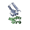
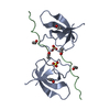
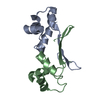
| Title | Mycobacterium tuberculosis HigA2 (Form I) | ||||||
 Components Components | Putative antitoxin HigA2 | ||||||
 Keywords Keywords | DNA BINDING PROTEIN / Antitoxin / HigA2 | ||||||
| Function / homology | Helix-turn-helix / Helix-turn-helix XRE-family like proteins / Cro/C1-type HTH domain profile. / Cro/C1-type helix-turn-helix domain / Lambda repressor-like, DNA-binding domain superfamily / DNA binding / Putative antitoxin HigA2 Function and homology information Function and homology information | ||||||
| Biological species |   Mycobacterium tuberculosis (bacteria) Mycobacterium tuberculosis (bacteria) | ||||||
| Method |  X-RAY DIFFRACTION / SYNCHROTRON / MOLECULAR REPLACEMENT / Resolution: 2.05 Å X-RAY DIFFRACTION / SYNCHROTRON / MOLECULAR REPLACEMENT / Resolution: 2.05 Å | ||||||
 Authors Authors | Kim, H.J. | ||||||
| Funding support |  Korea, Republic Of, 1items Korea, Republic Of, 1items
| ||||||
 Citation Citation | Journal: Iucrj / Year: 2021 Title: Chasing the structural diversity of the transcription regulator Mycobacterium tuberculosis HigA2. Authors: Richardson, W. / Kang, G.W. / Lee, H.J. / Kwon, K.M. / Kim, S. / Kim, H.J. | ||||||
| History |
|
- Structure visualization
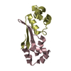
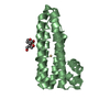
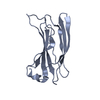
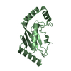
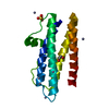
| Structure viewer | Molecule: MolmilJmol/JSmol |
|---|
- Downloads & links
-Download
| PDBx/mmCIF format | 7ewc.cif.gz | 66.6 KB | Display | PDBx/mmCIF format |
|---|---|---|---|---|
| PDB format | pdb7ewc.ent.gz | 48 KB | Display | PDB format |
| PDBx/mmJSON format | 7ewc.json.gz | Tree view | PDBx/mmJSON format | |
| Others |  Other downloads Other downloads |
-Validation report
| Summary document | 7ewc_validation.pdf.gz | 457.5 KB | Display | wwPDB validaton report |
|---|---|---|---|---|
| Full document | 7ewc_full_validation.pdf.gz | 462.3 KB | Display | |
| Data in XML | 7ewc_validation.xml.gz | 12.1 KB | Display | |
| Data in CIF | 7ewc_validation.cif.gz | 16.4 KB | Display | |
| Arichive directory | https://data.pdbj.org/pub/pdb/validation_reports/ew/7ewcftp://data.pdbj.org/pub/pdb/validation_reports/ew/7ewc | HTTPS FTP |
-Related structure data
| Related structure data | 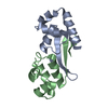 7ewdC  7eweC  1y7yS  2a6cS 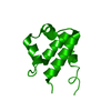 3b7hS 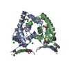 3kxaS S: Starting model for refinement C: citing same article ( |
|---|---|
| Similar structure data |

-Links
 PDBj
PDBj


- Assembly
| Deposited unit | 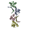
| ||||||||
|---|---|---|---|---|---|---|---|---|---|
| 1 | 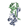
| ||||||||
| 2 | 
| ||||||||
| Unit cell |
|
-Components
| #1: Protein | Mass: 11309.927 Da / Num. of mol.: 4 Source method: isolated from a genetically manipulated source Source: (gene. exp.) Mycobacterium tuberculosis (strain ATCC 25618 / H37Rv) (bacteria)Gene: higA2, Rv2021c, RVBD_2021c, LH57_11010, P425_02092 / Production host: #2: Water | ChemComp-HOH / |  Mass: 18.015 Da / Num. of mol.: 54 / Source method: isolated from a natural source / Formula: H2O Mass: 18.015 Da / Num. of mol.: 54 / Source method: isolated from a natural source / Formula: H2O |
|---|
-Experimental details
-Experiment
| Experiment | Method: X-RAY DIFFRACTION / Number of used crystals: 1 |
|---|
- Sample preparation
| Crystal | Density Matthews: 1.73 Å3/Da / Density % sol: 28.89 % |
|---|---|
| Crystal grow | Temperature: 293 K / Method: vapor diffusion, hanging drop / Details: 11% (w/v) PEK 20000, 0.1M MES pH 6.5 |
-Data collection
| Diffraction | Mean temperature: 100 K / Serial crystal experiment: N |
|---|---|
| Diffraction source | Source: SYNCHROTRON / Site: PAL/PLS / Beamline: 5C (4A) / Wavelength: 0.98 Å |
| Detector | Type: ADSC QUANTUM 315 / Detector: CCD / Date: Jul 3, 2014 |
| Radiation | Protocol: SINGLE WAVELENGTH / Monochromatic (M) / Laue (L): M / Scattering type: x-ray |
| Radiation wavelength | Wavelength: 0.98 Å / Relative weight: 1 |
| Reflection | Resolution: 2.05→40 Å / Num. obs: 20447 / % possible obs: 99.8 % / Redundancy: 6 % / CC1/2: 0.99 / Rmerge(I) obs: 0.079 / Net I/σ(I): 29 |
| Reflection shell | Resolution: 2.05→2.09 Å / Num. unique obs: 969 / CC1/2: 0.984 |
- Processing
| Software |
| ||||||||||||||||||||||||||||||||||||||||||||||||||||||||||||
|---|---|---|---|---|---|---|---|---|---|---|---|---|---|---|---|---|---|---|---|---|---|---|---|---|---|---|---|---|---|---|---|---|---|---|---|---|---|---|---|---|---|---|---|---|---|---|---|---|---|---|---|---|---|---|---|---|---|---|---|---|---|
| Refinement | Method to determine structure: MOLECULAR REPLACEMENT Starting model: 1Y7Y, 3B7H, 3KXA, 2A6C Resolution: 2.05→35.22 Å / Cor.coef. Fo:Fc: 0.946 / Cor.coef. Fo:Fc free: 0.924 / SU B: 4.201 / SU ML: 0.116 / Cross valid method: THROUGHOUT / σ(F): 0 / ESU R: 0.194 / ESU R Free: 0.172 / Stereochemistry target values: MAXIMUM LIKELIHOOD Details: HYDROGENS HAVE BEEN ADDED IN THE RIDING POSITIONS U VALUES : REFINED INDIVIDUALLY
| ||||||||||||||||||||||||||||||||||||||||||||||||||||||||||||
| Solvent computation | Ion probe radii: 0.8 Å / Shrinkage radii: 0.8 Å / VDW probe radii: 1.2 Å / Solvent model: MASK | ||||||||||||||||||||||||||||||||||||||||||||||||||||||||||||
| Displacement parameters | Biso max: 95.22 Å2 / Biso mean: 35.407 Å2 / Biso min: 12.96 Å2
| ||||||||||||||||||||||||||||||||||||||||||||||||||||||||||||
| Refinement step | Cycle: final / Resolution: 2.05→35.22 Å
| ||||||||||||||||||||||||||||||||||||||||||||||||||||||||||||
| Refine LS restraints |
| ||||||||||||||||||||||||||||||||||||||||||||||||||||||||||||
| LS refinement shell | Resolution: 2.05→2.103 Å / Rfactor Rfree error: 0 / Total num. of bins used: 20
|
