 Movie
Movie Controller

[English] 日本語
 Yorodumi
- PDB-1y7y: High-resolution crystal structure of the restriction-modification... -
+ Open data

- Basic information
| Entry | Database: PDB / ID: 1y7y | ||||||
|---|---|---|---|---|---|---|---|
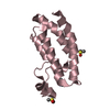
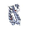
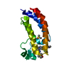
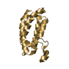
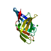
| Title | High-resolution crystal structure of the restriction-modification controller protein C.AhdI from Aeromonas hydrophila | ||||||
 Components Components | C.AhdI | ||||||
 Keywords Keywords | Transcription regulator / HELIX-TURN-HELIX / DNA-BINDING PROTEIN / TRANSCRIPTIONAL REGULATOR | ||||||
| Function / homology |  Function and homology information Function and homology information | ||||||
| Biological species |  Aeromonas hydrophila (bacteria) Aeromonas hydrophila (bacteria) | ||||||
| Method |  X-RAY DIFFRACTION / SYNCHROTRON / MOLECULAR REPLACEMENT / Resolution: 1.69 Å X-RAY DIFFRACTION / SYNCHROTRON / MOLECULAR REPLACEMENT / Resolution: 1.69 Å | ||||||
 Authors Authors | McGeehan, J.E. / Streeter, S.D. / Papapanagiotou, I. / Fox, G.C. / Kneale, G.G. | ||||||
 Citation Citation | Journal: J.Mol.Biol. / Year: 2005 Title: High-resolution crystal structure of the restriction-modification controller protein C.AhdI from Aeromonas hydrophila. Authors: McGeehan, J.E. / Streeter, S.D. / Papapanagiotou, I. / Fox, G.C. / Kneale, G.G. #1: Journal: Acta Crystallogr.,Sect.D / Year: 2004 Title: Crystallization and preliminary X-ray analysis of the controller protein C.AhdI from Aeromonas hydrophilia Authors: McGeehan, J.E. / Streeter, S. / Cooper, J.B. / Mohammed, F. / Fox, G.C. / Kneale, G.G. | ||||||
| History |
|
- Structure visualization
| Structure viewer | Molecule: MolmilJmol/JSmol |
|---|
- Downloads & links
-Download
| PDBx/mmCIF format | 1y7y.cif.gz | 41.6 KB | Display | PDBx/mmCIF format |
|---|---|---|---|---|
| PDB format | pdb1y7y.ent.gz | 29.8 KB | Display | PDB format |
| PDBx/mmJSON format | 1y7y.json.gz | Tree view | PDBx/mmJSON format | |
| Others |  Other downloads Other downloads |
-Validation report
| Arichive directory | https://data.pdbj.org/pub/pdb/validation_reports/y7/1y7yftp://data.pdbj.org/pub/pdb/validation_reports/y7/1y7y | HTTPS FTP |
|---|
-Related structure data
| Similar structure data |
|---|


-Links
 PDBj
PDBj


- Assembly
| Deposited unit | 
| ||||||||
|---|---|---|---|---|---|---|---|---|---|
| 1 |
| ||||||||
| Unit cell |
| ||||||||
| Details | THIS ENTRY CONTAINS THE CRYSTALLOGRAPHIC ASYMMETRIC UNIT WHICH CONSISTS OF 2 CHAINS WHICH MAKE UP THE BIOLOGICAL DIMER |
-Components
| #1: Protein | Mass: 8382.575 Da / Num. of mol.: 2 Source method: isolated from a genetically manipulated source Source: (gene. exp.) Aeromonas hydrophila (bacteria) / Plasmid: pET11a / Production host: #2: Water | ChemComp-HOH / |  Mass: 18.015 Da / Num. of mol.: 147 / Source method: isolated from a natural source / Formula: H2O Mass: 18.015 Da / Num. of mol.: 147 / Source method: isolated from a natural source / Formula: H2O |
|---|
-Experimental details
-Experiment
| Experiment | Method: X-RAY DIFFRACTION / Number of used crystals: 1 |
|---|
- Sample preparation
| Crystal | Density Matthews: 1.89 Å3/Da / Density % sol: 34.9 % |
|---|---|
| Crystal grow | Temperature: 295 K / Method: vapor diffusion, hanging drop / pH: 7 Details: PEG 6000, IMIDAZOLE-MALATE, 2-METHYL-2,4-PENTANEDIOL, pH 7.0, VAPOR DIFFUSION, HANGING DROP, temperature 295K |
-Data collection
| Diffraction | Mean temperature: 100 K |
|---|---|
| Diffraction source | Source: SYNCHROTRON / Site: ESRF  / Beamline: BM14 / Wavelength: 0.904989 Å / Beamline: BM14 / Wavelength: 0.904989 Å |
| Detector | Type: MARRESEARCH / Detector: CCD / Date: Jun 21, 2003 / Details: MIRRORS |
| Radiation | Monochromator: Si 111 CHANNEL / Protocol: SINGLE WAVELENGTH / Monochromatic (M) / Laue (L): M / Scattering type: x-ray |
| Radiation wavelength | Wavelength: 0.904989 Å / Relative weight: 1 |
| Reflection | Resolution: 1.66→45.57 Å / Num. all: 14937 / Num. obs: 14302 / % possible obs: 95.7 % / Redundancy: 9.2 % / Biso Wilson estimate: 18.8 Å2 / Rmerge(I) obs: 0.048 / Rsym value: 0.041 / Net I/σ(I): 8 |
| Reflection shell | Resolution: 1.66→1.75 Å / Redundancy: 3 % / Rmerge(I) obs: 0.215 / Mean I/σ(I) obs: 4.1 / Num. unique all: 1561 / Rsym value: 0.181 / % possible all: 95.7 |
- Processing
| Software |
| |||||||||||||||||||||||||
|---|---|---|---|---|---|---|---|---|---|---|---|---|---|---|---|---|---|---|---|---|---|---|---|---|---|---|
| Refinement | Method to determine structure: MOLECULAR REPLACEMENT Starting model: Mutant I58M of C.AhdI, SeMet MAD to 2.5A Resolution: 1.69→18.4 Å / Cross valid method: THROUGHOUT / Stereochemistry target values: Engh & Huber
| |||||||||||||||||||||||||
| Refinement step | Cycle: LAST / Resolution: 1.69→18.4 Å
| |||||||||||||||||||||||||
| Refine LS restraints |
|
