 Movie
Movie Controller

[English] 日本語
 Yorodumi
- PDB-7ess: Structure-guided studies of the Holliday junction resolvase RuvX ... -
+ Open data

- Basic information
| Entry | Database: PDB / ID: 7ess | |||||||||
|---|---|---|---|---|---|---|---|---|---|---|
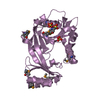
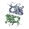
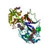
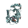
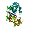
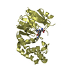
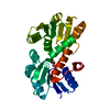
| Title | Structure-guided studies of the Holliday junction resolvase RuvX provide novel insights into ATP-stimulated cleavage of branched DNA and RNA substrates | |||||||||
 Components Components | Putative pre-16S rRNA nuclease | |||||||||
 Keywords Keywords | DNA BINDING PROTEIN / Holliday junction / Holliday junction resolvases / ATPase | |||||||||
| Function / homology |  Function and homology information Function and homology informationrRNA 5'-end processing / nuclease activity / Hydrolases; Acting on ester bonds / cytosol Similarity search - Function | |||||||||
| Biological species |  Mycobacterium tuberculosis H37Rv (bacteria) Mycobacterium tuberculosis H37Rv (bacteria) | |||||||||
| Method |  X-RAY DIFFRACTION / MOLECULAR REPLACEMENT / Resolution: 1.93 Å X-RAY DIFFRACTION / MOLECULAR REPLACEMENT / Resolution: 1.93 Å | |||||||||
 Authors Authors | Thakur, M. / Mohan, D. / Singh, A.K. / Agarwal, A. / Gopal, B. / Muniyappa, K. | |||||||||
| Funding support |  India, 2items India, 2items
| |||||||||
 Citation Citation | Journal: J.Mol.Biol. / Year: 2021 Title: Novel insights into ATP-Stimulated Cleavage of branched DNA and RNA Substrates through Structure-Guided Studies of the Holliday Junction Resolvase RuvX. Authors: Thakur, M. / Mohan, D. / Singh, A.K. / Agarwal, A. / Gopal, B. / Muniyappa, K. | |||||||||
| History |
|
- Structure visualization
| Structure viewer | Molecule: MolmilJmol/JSmol |
|---|
- Downloads & links
-Download
| PDBx/mmCIF format | 7ess.cif.gz | 73.3 KB | Display | PDBx/mmCIF format |
|---|---|---|---|---|
| PDB format | pdb7ess.ent.gz | 52.8 KB | Display | PDB format |
| PDBx/mmJSON format | 7ess.json.gz | Tree view | PDBx/mmJSON format | |
| Others |  Other downloads Other downloads |
-Validation report
| Summary document | 7ess_validation.pdf.gz | 436 KB | Display | wwPDB validaton report |
|---|---|---|---|---|
| Full document | 7ess_full_validation.pdf.gz | 437.5 KB | Display | |
| Data in XML | 7ess_validation.xml.gz | 14.6 KB | Display | |
| Data in CIF | 7ess_validation.cif.gz | 20.8 KB | Display | |
| Arichive directory | https://data.pdbj.org/pub/pdb/validation_reports/es/7essftp://data.pdbj.org/pub/pdb/validation_reports/es/7ess | HTTPS FTP |
-Related structure data
| Related structure data |  1vhxS S: Starting model for refinement |
|---|---|
| Similar structure data |


-Links
 PDBj
PDBj- Assembly
| Deposited unit | 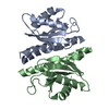
| ||||||||
|---|---|---|---|---|---|---|---|---|---|
| 1 |
| ||||||||
| Unit cell |
|
-Components
| #1: Protein | Mass: 18092.498 Da / Num. of mol.: 2 Source method: isolated from a genetically manipulated source Source: (gene. exp.) Mycobacterium tuberculosis H37Rv (bacteria)Strain: H37Rv / Gene: Rv2554c, MTCY159.02 / Production host: References: UniProt: P9WGV7, Hydrolases; Acting on ester bonds #2: Water | ChemComp-HOH / |  Mass: 18.015 Da / Num. of mol.: 185 / Source method: isolated from a natural source / Formula: H2O Mass: 18.015 Da / Num. of mol.: 185 / Source method: isolated from a natural source / Formula: H2O |
|---|
-Experimental details
-Experiment
| Experiment | Method: X-RAY DIFFRACTION / Number of used crystals: 1 |
|---|
- Sample preparation
| Crystal | Density Matthews: 1.83 Å3/Da / Density % sol: 32.73 % |
|---|---|
| Crystal grow | Temperature: 300 K / Method: batch mode / pH: 7 Details: 0.1 M Bis-Tris propane, pH 7.0 and 2.4 M sodium malonate, pH 7.0 PH range: 7 |
-Data collection
| Diffraction | Mean temperature: 100 K / Serial crystal experiment: N | |||||||||||||||||||||||||||||||||||||||||||||||||||||||||||||||||||||||||||||||||||||||||||||||||||
|---|---|---|---|---|---|---|---|---|---|---|---|---|---|---|---|---|---|---|---|---|---|---|---|---|---|---|---|---|---|---|---|---|---|---|---|---|---|---|---|---|---|---|---|---|---|---|---|---|---|---|---|---|---|---|---|---|---|---|---|---|---|---|---|---|---|---|---|---|---|---|---|---|---|---|---|---|---|---|---|---|---|---|---|---|---|---|---|---|---|---|---|---|---|---|---|---|---|---|---|---|
| Diffraction source | Source: ROTATING ANODE / Type: RIGAKU FR-E SUPERBRIGHT / Wavelength: 1.5418 Å | |||||||||||||||||||||||||||||||||||||||||||||||||||||||||||||||||||||||||||||||||||||||||||||||||||
| Detector | Type: RIGAKU RAXIS / Detector: IMAGE PLATE / Date: Mar 28, 2021 | |||||||||||||||||||||||||||||||||||||||||||||||||||||||||||||||||||||||||||||||||||||||||||||||||||
| Radiation | Protocol: SINGLE WAVELENGTH / Monochromatic (M) / Laue (L): M / Scattering type: x-ray | |||||||||||||||||||||||||||||||||||||||||||||||||||||||||||||||||||||||||||||||||||||||||||||||||||
| Radiation wavelength | Wavelength: 1.5418 Å / Relative weight: 1 | |||||||||||||||||||||||||||||||||||||||||||||||||||||||||||||||||||||||||||||||||||||||||||||||||||
| Reflection | Resolution: 1.93→53.477 Å / Num. obs: 19756 / % possible obs: 99.3 % / Redundancy: 4.7 % / Rpim(I) all: 0.034 / Rrim(I) all: 0.075 / Rsym value: 0.067 / Net I/av σ(I): 8.1 / Net I/σ(I): 13.2 | |||||||||||||||||||||||||||||||||||||||||||||||||||||||||||||||||||||||||||||||||||||||||||||||||||
| Reflection shell | Diffraction-ID: 1
|
- Processing
| Software |
| ||||||||||||||||||||||||||||||||||||||||||||||||||||||||||||
|---|---|---|---|---|---|---|---|---|---|---|---|---|---|---|---|---|---|---|---|---|---|---|---|---|---|---|---|---|---|---|---|---|---|---|---|---|---|---|---|---|---|---|---|---|---|---|---|---|---|---|---|---|---|---|---|---|---|---|---|---|---|
| Refinement | Method to determine structure: MOLECULAR REPLACEMENT Starting model: 1VHX Resolution: 1.93→52.95 Å / Cor.coef. Fo:Fc: 0.949 / Cor.coef. Fo:Fc free: 0.927 / Cross valid method: THROUGHOUT / σ(F): 0 / ESU R: 0.182 / ESU R Free: 0.159 / Stereochemistry target values: MAXIMUM LIKELIHOOD Details: HYDROGENS HAVE BEEN ADDED IN THE RIDING POSITIONS U VALUES : REFINED INDIVIDUALLY
| ||||||||||||||||||||||||||||||||||||||||||||||||||||||||||||
| Solvent computation | Ion probe radii: 0.8 Å / Shrinkage radii: 0.8 Å / VDW probe radii: 1.2 Å / Solvent model: MASK | ||||||||||||||||||||||||||||||||||||||||||||||||||||||||||||
| Displacement parameters | Biso max: 107.24 Å2 / Biso mean: 22.757 Å2 / Biso min: 10.5 Å2
| ||||||||||||||||||||||||||||||||||||||||||||||||||||||||||||
| Refinement step | Cycle: final / Resolution: 1.93→52.95 Å
| ||||||||||||||||||||||||||||||||||||||||||||||||||||||||||||
| Refine LS restraints |
| ||||||||||||||||||||||||||||||||||||||||||||||||||||||||||||
| LS refinement shell | Resolution: 1.931→1.981 Å / Rfactor Rfree error: 0 / Total num. of bins used: 20
|
