 Movie
Movie Controller


+ Open data

- Basic information
| Entry | Database: PDB / ID: 2pt2 | ||||||
|---|---|---|---|---|---|---|---|
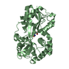
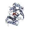
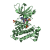
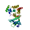
| Title | Structure of FutA1 with Iron(II) | ||||||
 Components Components | Iron transport protein | ||||||
 Keywords Keywords | METAL TRANSPORT / C-clamp / iron-binding protein / solute-binding protein / periplasmic binding protein / ABC transporter | ||||||
| Function / homology |  Function and homology information Function and homology informationplasma membrane-derived thylakoid membrane / iron ion transport / metal ion binding Similarity search - Function | ||||||
| Biological species |  | ||||||
| Method |  X-RAY DIFFRACTION / SYNCHROTRON / MOLECULAR REPLACEMENT / Resolution: 2 Å X-RAY DIFFRACTION / SYNCHROTRON / MOLECULAR REPLACEMENT / Resolution: 2 Å | ||||||
 Authors Authors | Koropatkin, N.M. / Randich, A.M. / Bhattachryya-Pakrasi, M. / Pakrasi, H.B. / Smith, T.J. | ||||||
 Citation Citation | Journal: J.Biol.Chem. / Year: 2007 Title: The structure of the iron-binding protein, FutA1, from Synechocystis 6803. Authors: Koropatkin, N. / Randich, A.M. / Bhattacharyya-Pakrasi, M. / Pakrasi, H.B. / Smith, T.J. | ||||||
| History |
|
- Structure visualization
| Structure viewer | Molecule: MolmilJmol/JSmol |
|---|
- Downloads & links
-Download
| PDBx/mmCIF format | 2pt2.cif.gz | 83.6 KB | Display | PDBx/mmCIF format |
|---|---|---|---|---|
| PDB format | pdb2pt2.ent.gz | 60.2 KB | Display | PDB format |
| PDBx/mmJSON format | 2pt2.json.gz | Tree view | PDBx/mmJSON format | |
| Others |  Other downloads Other downloads |
-Validation report
| Arichive directory | https://data.pdbj.org/pub/pdb/validation_reports/pt/2pt2ftp://data.pdbj.org/pub/pdb/validation_reports/pt/2pt2 | HTTPS FTP |
|---|
-Related structure data
| Related structure data | 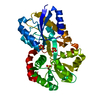 2pt1SC  3f11C S: Starting model for refinement C: citing same article ( |
|---|---|
| Similar structure data |


-Links
 PDBj
PDBj


- Assembly
| Deposited unit | 
| ||||||||
|---|---|---|---|---|---|---|---|---|---|
| 1 |
| ||||||||
| Unit cell |
|
-Components
| #1: Protein | Mass: 36606.691 Da / Num. of mol.: 1 Source method: isolated from a genetically manipulated source Source: (gene. exp.) | ||||
|---|---|---|---|---|---|
| #2: Chemical | ChemComp-FE2 /   Mass: 55.845 Da / Num. of mol.: 1 / Source method: obtained synthetically / Formula: Fe Mass: 55.845 Da / Num. of mol.: 1 / Source method: obtained synthetically / Formula: Fe | ||||
| #3: Chemical | ChemComp-SO4 /   Mass: 96.063 Da / Num. of mol.: 4 / Source method: obtained synthetically / Formula: SO4 Mass: 96.063 Da / Num. of mol.: 4 / Source method: obtained synthetically / Formula: SO4#4: Chemical | ChemComp-EDO / |   Mass: 62.068 Da / Num. of mol.: 1 / Source method: obtained synthetically / Formula: C2H6O2 Mass: 62.068 Da / Num. of mol.: 1 / Source method: obtained synthetically / Formula: C2H6O2#5: Water | ChemComp-HOH / |  Mass: 18.015 Da / Num. of mol.: 318 / Source method: isolated from a natural source / Formula: H2O Mass: 18.015 Da / Num. of mol.: 318 / Source method: isolated from a natural source / Formula: H2O |
-Experimental details
-Experiment
| Experiment | Method: X-RAY DIFFRACTION / Number of used crystals: 1 |
|---|
- Sample preparation
| Crystal | Density Matthews: 2.16 Å3/Da / Density % sol: 43.15 % |
|---|---|
| Crystal grow | Temperature: 298 K / Method: batch seeding / pH: 8 Details: 100mM HEPPS, 2.2-2.8 ammonium sulfate, , pH 8.0, batch seeding, temperature 298K |
-Data collection
| Diffraction | Mean temperature: 100 K |
|---|---|
| Diffraction source | Source: SYNCHROTRON / Site: APS  / Beamline: 19-ID / Wavelength: 0.97167 Å / Beamline: 19-ID / Wavelength: 0.97167 Å |
| Detector | Type: CUSTOM-MADE / Detector: CCD / Date: Nov 16, 2006 / Details: mirrors |
| Radiation | Monochromator: si 111 / Protocol: SINGLE WAVELENGTH / Monochromatic (M) / Laue (L): M / Scattering type: x-ray |
| Radiation wavelength | Wavelength: 0.97167 Å / Relative weight: 1 |
| Reflection | Resolution: 2→50 Å / Num. all: 20599 / Num. obs: 20599 / % possible obs: 92.8 % / Observed criterion σ(F): 0 / Observed criterion σ(I): 0 / Redundancy: 4.8 % / Rsym value: 0.113 / Net I/σ(I): 43.7 |
| Reflection shell | Resolution: 2→2.02 Å / Redundancy: 3.4 % / Mean I/σ(I) obs: 20.8 / Num. unique all: 505 / Rsym value: 0.145 / % possible all: 86.8 |
- Processing
| Software |
| ||||||||||||||||||||||||||||
|---|---|---|---|---|---|---|---|---|---|---|---|---|---|---|---|---|---|---|---|---|---|---|---|---|---|---|---|---|---|
| Refinement | Method to determine structure: MOLECULAR REPLACEMENT Starting model: FutA1; entry 2PT1 Resolution: 2→50 Å / Cross valid method: THROUGHOUT / σ(F): 0 / σ(I): 0 / Stereochemistry target values: Engh & Huber
| ||||||||||||||||||||||||||||
| Solvent computation | Bsol: 40.887 Å2 | ||||||||||||||||||||||||||||
| Displacement parameters | Biso mean: 19.348 Å2
| ||||||||||||||||||||||||||||
| Refinement step | Cycle: LAST / Resolution: 2→50 Å
| ||||||||||||||||||||||||||||
| Refine LS restraints |
| ||||||||||||||||||||||||||||
| LS refinement shell | Resolution: 2→2.02 Å
| ||||||||||||||||||||||||||||
| Xplor file |
|
