 Movie
Movie Controller

[English] 日本語
 Yorodumi
- PDB-7emg: Carbonyl Reductase Variant 4 (R123C/L209P/F183Y/V61K) from Serrat... -
+ Open data

- Basic information
| Entry | Database: PDB / ID: 7emg | ||||||
|---|---|---|---|---|---|---|---|
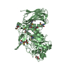
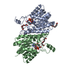
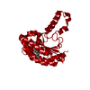
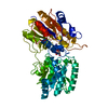
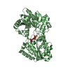
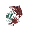
| Title | Carbonyl Reductase Variant 4 (R123C/L209P/F183Y/V61K) from Serratia marcescens complexed with NADP+ | ||||||
 Components Components | 3-oxoacyl-[acyl-carrier-protein] reductase | ||||||
 Keywords Keywords | OXIDOREDUCTASE / NADP+ complex / carbonyl reductase | ||||||
| Function / homology |  Function and homology information Function and homology information: / : / 3-oxoacyl-[acyl-carrier-protein] reductase (NADPH) activity / 3-oxoacyl-[acyl-carrier-protein] reductase / NAD binding / fatty acid biosynthetic process Similarity search - Function | ||||||
| Biological species |  Serratia marcescens (bacteria) Serratia marcescens (bacteria) | ||||||
| Method |  X-RAY DIFFRACTION / SYNCHROTRON / MOLECULAR REPLACEMENT / Resolution: 2.449 Å X-RAY DIFFRACTION / SYNCHROTRON / MOLECULAR REPLACEMENT / Resolution: 2.449 Å | ||||||
 Authors Authors | Zheng, Y.C. / Wang, T. / Bai, Y.P. | ||||||
 Citation Citation | Journal: Chem.Commun.(Camb.) / Year: 2021 Title: Stereoselective synthesis of chiral delta-lactones via an engineered carbonyl reductase. Authors: Wang, T. / Zhang, X.Y. / Zheng, Y.C. / Bai, Y.P. | ||||||
| History |
|
- Structure visualization
| Structure viewer | Molecule: MolmilJmol/JSmol |
|---|
- Downloads & links
-Download
| PDBx/mmCIF format | 7emg.cif.gz | 104.8 KB | Display | PDBx/mmCIF format |
|---|---|---|---|---|
| PDB format | pdb7emg.ent.gz | 77.7 KB | Display | PDB format |
| PDBx/mmJSON format | 7emg.json.gz | Tree view | PDBx/mmJSON format | |
| Others |  Other downloads Other downloads |
-Validation report
| Summary document | 7emg_validation.pdf.gz | 1.1 MB | Display | wwPDB validaton report |
|---|---|---|---|---|
| Full document | 7emg_full_validation.pdf.gz | 1.1 MB | Display | |
| Data in XML | 7emg_validation.xml.gz | 20.4 KB | Display | |
| Data in CIF | 7emg_validation.cif.gz | 26.7 KB | Display | |
| Arichive directory | https://data.pdbj.org/pub/pdb/validation_reports/em/7emgftp://data.pdbj.org/pub/pdb/validation_reports/em/7emg | HTTPS FTP |
-Related structure data
| Related structure data | 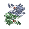 1q7cS S: Starting model for refinement |
|---|---|
| Similar structure data |


-Links
 PDBj
PDBj


- Assembly
| Deposited unit | 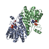
| ||||||||
|---|---|---|---|---|---|---|---|---|---|
| 1 |
| ||||||||
| Unit cell |
|
-Components
| #1: Protein | Mass: 26624.428 Da / Num. of mol.: 2 / Mutation: V61K, R123C, F183Y, L209P Source method: isolated from a genetically manipulated source Source: (gene. exp.) Serratia marcescens (bacteria)Gene: fabG, A8A12_06940, AR325_11665, BVG93_06205, G3M86_05635, G3M87_05635, G3M88_05635, G3M89_05635, GV243_09700, HMI62_09690 Production host: References: UniProt: A0A0G8B235, 3-oxoacyl-[acyl-carrier-protein] reductase #2: Chemical |   Mass: 745.421 Da / Num. of mol.: 2 / Source method: obtained synthetically / Formula: C21H30N7O17P3 Mass: 745.421 Da / Num. of mol.: 2 / Source method: obtained synthetically / Formula: C21H30N7O17P3#3: Chemical | ChemComp-EDO / |   Mass: 62.068 Da / Num. of mol.: 1 / Source method: obtained synthetically / Formula: C2H6O2 Mass: 62.068 Da / Num. of mol.: 1 / Source method: obtained synthetically / Formula: C2H6O2#4: Water | ChemComp-HOH / |  Mass: 18.015 Da / Num. of mol.: 58 / Source method: isolated from a natural source / Formula: H2O Mass: 18.015 Da / Num. of mol.: 58 / Source method: isolated from a natural source / Formula: H2OHas ligand of interest | N | |
|---|
-Experimental details
-Experiment
| Experiment | Method: X-RAY DIFFRACTION / Number of used crystals: 1 |
|---|
- Sample preparation
| Crystal | Density Matthews: 2.18 Å3/Da / Density % sol: 43.46 % |
|---|---|
| Crystal grow | Temperature: 291 K / Method: vapor diffusion, sitting drop Details: 0.1M Sodium formate; 0.1M Ammonium acetate; 0.1M Sodium citrate tribasic dihydrate; 0.1M Potassium sodium tartrate tetrahydrate; 0.1M Sodium oxamate; 0.1M Imidazole; 0.1M MES monohydrate ...Details: 0.1M Sodium formate; 0.1M Ammonium acetate; 0.1M Sodium citrate tribasic dihydrate; 0.1M Potassium sodium tartrate tetrahydrate; 0.1M Sodium oxamate; 0.1M Imidazole; 0.1M MES monohydrate (acid); pH 6.5; 12.5% v/v MPD; 12.5% PEG 1000; 12.5% w/v PEG 3350 PH range: 6.5-7.5 |
-Data collection
| Diffraction | Mean temperature: 100 K / Serial crystal experiment: N |
|---|---|
| Diffraction source | Source: SYNCHROTRON / Site: SSRF  / Beamline: BL19U1 / Wavelength: 0.978 Å / Beamline: BL19U1 / Wavelength: 0.978 Å |
| Detector | Type: DECTRIS PILATUS3 6M / Detector: PIXEL / Date: Nov 28, 2019 |
| Radiation | Protocol: SINGLE WAVELENGTH / Monochromatic (M) / Laue (L): M / Scattering type: x-ray |
| Radiation wavelength | Wavelength: 0.978 Å / Relative weight: 1 |
| Reflection | Resolution: 2.449→50 Å / Num. obs: 17156 / % possible obs: 99 % / Redundancy: 12.8 % / Biso Wilson estimate: 33.5 Å2 / CC1/2: 0.969 / CC star: 0.992 / Χ2: 0.878 / Net I/σ(I): 15 |
| Reflection shell | Resolution: 2.449→2.49 Å / Mean I/σ(I) obs: 4.1 / Num. unique obs: 849 / CC1/2: 0.954 / CC star: 0.988 / Χ2: 1.016 |
- Processing
| Software |
| ||||||||||||||||||||||||||||||||||||
|---|---|---|---|---|---|---|---|---|---|---|---|---|---|---|---|---|---|---|---|---|---|---|---|---|---|---|---|---|---|---|---|---|---|---|---|---|---|
| Refinement | Method to determine structure: MOLECULAR REPLACEMENT Starting model: 1q7c Resolution: 2.449→42.596 Å / SU ML: 0.24 / Cross valid method: THROUGHOUT / σ(F): 1.34 / Phase error: 27.31 / Stereochemistry target values: ML
| ||||||||||||||||||||||||||||||||||||
| Solvent computation | Shrinkage radii: 0.9 Å / VDW probe radii: 1.11 Å / Solvent model: FLAT BULK SOLVENT MODEL | ||||||||||||||||||||||||||||||||||||
| Displacement parameters | Biso max: 115.79 Å2 / Biso mean: 35.7916 Å2 / Biso min: 15.51 Å2 | ||||||||||||||||||||||||||||||||||||
| Refinement step | Cycle: final / Resolution: 2.449→42.596 Å
| ||||||||||||||||||||||||||||||||||||
| Refine LS restraints |
| ||||||||||||||||||||||||||||||||||||
| LS refinement shell | Refine-ID: X-RAY DIFFRACTION / Rfactor Rfree error: 0
|
