 Movie
Movie Controller

[English] 日本語
 Yorodumi
- PDB-7ekz: Structural and functional insights into Hydra Actinoporin-Like To... -
+ Open data

- Basic information
| Entry | Database: PDB / ID: 7ekz | |||||||||
|---|---|---|---|---|---|---|---|---|---|---|
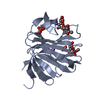
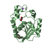
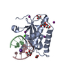
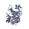
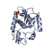
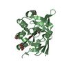
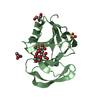
| Title | Structural and functional insights into Hydra Actinoporin-Like Toxin 1 (HALT-1) | |||||||||
 Components Components | HALT-1 | |||||||||
 Keywords Keywords | TOXIN / Hydra / actinoporin / HALT-1 / pore forming toxin | |||||||||
| Function / homology | FORMIC ACID Function and homology information Function and homology information | |||||||||
| Biological species |  | |||||||||
| Method |  X-RAY DIFFRACTION / MOLECULAR REPLACEMENT / Resolution: 1.43 Å X-RAY DIFFRACTION / MOLECULAR REPLACEMENT / Resolution: 1.43 Å | |||||||||
 Authors Authors | Ker, D.S. / Sha, X.H. / Jonet, M.A. / Hwang, J.S. / Ng, C.L. | |||||||||
| Funding support |  Malaysia, 2items Malaysia, 2items
| |||||||||
 Citation Citation | Journal: Sci Rep / Year: 2021 Title: Structural and functional analysis of Hydra Actinoporin-Like Toxin 1 (HALT-1). Authors: Ker, D.S. / Sha, H.X. / Jonet, M.A. / Hwang, J.S. / Ng, C.L. | |||||||||
| History |
|
- Structure visualization
| Structure viewer | Molecule: MolmilJmol/JSmol |
|---|
- Downloads & links
-Download
| PDBx/mmCIF format | 7ekz.cif.gz | 98.8 KB | Display | PDBx/mmCIF format |
|---|---|---|---|---|
| PDB format | pdb7ekz.ent.gz | 72.2 KB | Display | PDB format |
| PDBx/mmJSON format | 7ekz.json.gz | Tree view | PDBx/mmJSON format | |
| Others |  Other downloads Other downloads |
-Validation report
| Arichive directory | https://data.pdbj.org/pub/pdb/validation_reports/ek/7ekzftp://data.pdbj.org/pub/pdb/validation_reports/ek/7ekz | HTTPS FTP |
|---|
-Related structure data
| Related structure data |  1gwyS S: Starting model for refinement |
|---|---|
| Similar structure data |


-Links
 PDBj
PDBj- Assembly
| Deposited unit | 
| ||||||||
|---|---|---|---|---|---|---|---|---|---|
| 1 |
| ||||||||
| Unit cell |
|
-Components
| #1: Protein | Mass: 20758.477 Da / Num. of mol.: 1 Source method: isolated from a genetically manipulated source Source: (gene. exp.)  | ||||||||
|---|---|---|---|---|---|---|---|---|---|
| #2: Chemical |   Mass: 46.025 Da / Num. of mol.: 3 / Source method: obtained synthetically / Formula: CH2O2 Mass: 46.025 Da / Num. of mol.: 3 / Source method: obtained synthetically / Formula: CH2O2#3: Chemical |   Mass: 92.094 Da / Num. of mol.: 3 / Source method: obtained synthetically / Formula: C3H8O3 Mass: 92.094 Da / Num. of mol.: 3 / Source method: obtained synthetically / Formula: C3H8O3#4: Water | ChemComp-HOH / |  Mass: 18.015 Da / Num. of mol.: 345 / Source method: isolated from a natural source / Formula: H2O Mass: 18.015 Da / Num. of mol.: 345 / Source method: isolated from a natural source / Formula: H2OHas ligand of interest | N | Has protein modification | Y | |
-Experimental details
-Experiment
| Experiment | Method: X-RAY DIFFRACTION / Number of used crystals: 1 |
|---|
- Sample preparation
| Crystal | Density Matthews: 2.17 Å3/Da / Density % sol: 43.28 % |
|---|---|
| Crystal grow | Temperature: 293.15 K / Method: vapor diffusion, hanging drop / pH: 7.5 Details: 0.1 M sodium formate, 22% w/v polyethylene glycol 3550 |
-Data collection
| Diffraction | Mean temperature: 100 K / Serial crystal experiment: N |
|---|---|
| Diffraction source | Source: ROTATING ANODE / Type: RIGAKU MICROMAX-007 HF / Wavelength: 1.54187 Å |
| Detector | Type: DECTRIS PILATUS 200K / Detector: PIXEL / Date: Aug 29, 2018 |
| Radiation | Protocol: SINGLE WAVELENGTH / Monochromatic (M) / Laue (L): M / Scattering type: x-ray |
| Radiation wavelength | Wavelength: 1.54187 Å / Relative weight: 1 |
| Reflection | Resolution: 1.43→23.55 Å / Num. obs: 33402 / % possible obs: 98.2 % / Observed criterion σ(I): 2 / Redundancy: 4.8 % / CC1/2: 0.983 / Rmerge(I) obs: 0.147 / Rpim(I) all: 0.068 / Rrim(I) all: 0.163 / Net I/σ(I): 5.2 |
| Reflection shell | Resolution: 1.43→1.45 Å / Redundancy: 3 % / Rmerge(I) obs: 0.419 / Mean I/σ(I) obs: 2 / Num. unique obs: 1577 / CC1/2: 0.774 / Rpim(I) all: 0.276 / Rrim(I) all: 0.506 / % possible all: 95.2 |
- Processing
| Software |
| |||||||||||||||||||||||||||||||||||||||||||||||||||||||||||||||||||||||||||
|---|---|---|---|---|---|---|---|---|---|---|---|---|---|---|---|---|---|---|---|---|---|---|---|---|---|---|---|---|---|---|---|---|---|---|---|---|---|---|---|---|---|---|---|---|---|---|---|---|---|---|---|---|---|---|---|---|---|---|---|---|---|---|---|---|---|---|---|---|---|---|---|---|---|---|---|---|
| Refinement | Method to determine structure: MOLECULAR REPLACEMENT Starting model: 1GWY Resolution: 1.43→23.55 Å / Cor.coef. Fo:Fc: 0.957 / Cor.coef. Fo:Fc free: 0.919 / SU B: 3.009 / SU ML: 0.052 / Cross valid method: THROUGHOUT / σ(F): 0 / ESU R: 0.08 / ESU R Free: 0.077 / Stereochemistry target values: MAXIMUM LIKELIHOOD Details: HYDROGENS HAVE BEEN ADDED IN THE RIDING POSITIONS U VALUES : WITH TLS ADDED
| |||||||||||||||||||||||||||||||||||||||||||||||||||||||||||||||||||||||||||
| Solvent computation | Ion probe radii: 0.8 Å / Shrinkage radii: 0.8 Å / VDW probe radii: 1.2 Å / Solvent model: MASK | |||||||||||||||||||||||||||||||||||||||||||||||||||||||||||||||||||||||||||
| Displacement parameters | Biso max: 174.23 Å2 / Biso mean: 11.024 Å2 / Biso min: 2.93 Å2
| |||||||||||||||||||||||||||||||||||||||||||||||||||||||||||||||||||||||||||
| Refinement step | Cycle: final / Resolution: 1.43→23.55 Å
| |||||||||||||||||||||||||||||||||||||||||||||||||||||||||||||||||||||||||||
| Refine LS restraints |
| |||||||||||||||||||||||||||||||||||||||||||||||||||||||||||||||||||||||||||
| LS refinement shell | Resolution: 1.43→1.467 Å / Rfactor Rfree error: 0 / Total num. of bins used: 20
| |||||||||||||||||||||||||||||||||||||||||||||||||||||||||||||||||||||||||||
| Refinement TLS params. | Method: refined / Origin x: 2.2179 Å / Origin y: -15.9493 Å / Origin z: -11.4596 Å
|
