 Movie
Movie Controller

[English] 日本語
 Yorodumi
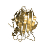
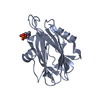
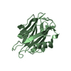
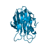
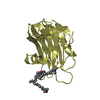
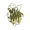
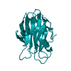
- PDB-1gwy: Crystal structure of the water-soluble state of the pore-forming ... -
+ Open data

- Basic information
| Entry | Database: PDB / ID: 1gwy | ||||||
|---|---|---|---|---|---|---|---|
| Title | Crystal structure of the water-soluble state of the pore-forming cytolysin Sticholysin II | ||||||
 Components Components | STICHOLYSIN II | ||||||
 Keywords Keywords | CYTOLYSIN / PORE-FORMING TOXIN / HEMOLYSIS / CNIDOCYST | ||||||
| Function / homology |  Function and homology information Function and homology informationnematocyst / cytolysis in another organism / pore complex assembly / other organism cell membrane / pore complex / monoatomic cation transport / toxin activity / channel activity / extracellular region / identical protein binding Similarity search - Function | ||||||
| Biological species |  STOICHACTIS HELIANTHUS (sea anemone) STOICHACTIS HELIANTHUS (sea anemone) | ||||||
| Method |  X-RAY DIFFRACTION / SYNCHROTRON / MOLECULAR REPLACEMENT / Resolution: 1.71 Å X-RAY DIFFRACTION / SYNCHROTRON / MOLECULAR REPLACEMENT / Resolution: 1.71 Å | ||||||
 Authors Authors | Mancheno, J.M. / Martin-Benito, J. / Martinez-Ripoll, M. / Gavilanes, J.G. / Hermoso, J.A. | ||||||
 Citation Citation | Journal: Structure / Year: 2003 Title: Crystal and Electron Microscopy Structures of Sticholysin II Actinoporin Reveal Insights Into the Mechanism of Membrane Pore Formation Authors: Mancheno, J.M. / Martin-Benito, J. / Martinez-Ripoll, M. / Gavilanes, J.G. / Hermoso, J.A. | ||||||
| History |
|
- Structure visualization
| Structure viewer | Molecule: MolmilJmol/JSmol |
|---|
- Downloads & links
-Download
| PDBx/mmCIF format | 1gwy.cif.gz | 87.8 KB | Display | PDBx/mmCIF format |
|---|---|---|---|---|
| PDB format | pdb1gwy.ent.gz | 67 KB | Display | PDB format |
| PDBx/mmJSON format | 1gwy.json.gz | Tree view | PDBx/mmJSON format | |
| Others |  Other downloads Other downloads |
-Validation report
| Arichive directory | https://data.pdbj.org/pub/pdb/validation_reports/gw/1gwyftp://data.pdbj.org/pub/pdb/validation_reports/gw/1gwy | HTTPS FTP |
|---|
-Related structure data
| Related structure data |  1o71C  1o72C  1iazS C: citing same article ( S: Starting model for refinement |
|---|---|
| Similar structure data |


-Links
 PDBj
PDBj- Assembly
| Deposited unit | 
| ||||||||
|---|---|---|---|---|---|---|---|---|---|
| 1 | 
| ||||||||
| 2 | 
| ||||||||
| Unit cell |
|
-Components
| #1: Protein | Mass: 19302.844 Da / Num. of mol.: 2 / Source method: isolated from a natural source / Source: (natural) STOICHACTIS HELIANTHUS (sea anemone) / References: UniProt: P07845#2: Chemical | ChemComp-SO4 /   Mass: 96.063 Da / Num. of mol.: 8 / Source method: obtained synthetically / Formula: SO4 Mass: 96.063 Da / Num. of mol.: 8 / Source method: obtained synthetically / Formula: SO4#3: Water | ChemComp-HOH / |  Mass: 18.015 Da / Num. of mol.: 373 / Source method: isolated from a natural source / Formula: H2O Mass: 18.015 Da / Num. of mol.: 373 / Source method: isolated from a natural source / Formula: H2O |
|---|
-Experimental details
-Experiment
| Experiment | Method: X-RAY DIFFRACTION / Number of used crystals: 1 |
|---|
- Sample preparation
| Crystal | Density Matthews: 2.13 Å3/Da / Density % sol: 42 % | ||||||||||||||||||||||||||||||||||||
|---|---|---|---|---|---|---|---|---|---|---|---|---|---|---|---|---|---|---|---|---|---|---|---|---|---|---|---|---|---|---|---|---|---|---|---|---|---|
| Crystal grow | pH: 7.5 / Details: pH 7.50 | ||||||||||||||||||||||||||||||||||||
| Crystal grow | *PLUS pH: 7 / Method: vapor diffusion, hanging drop | ||||||||||||||||||||||||||||||||||||
| Components of the solutions | *PLUS
|
-Data collection
| Diffraction | Mean temperature: 120 K |
|---|---|
| Diffraction source | Source: SYNCHROTRON / Site: ESRF  / Beamline: BM14 / Wavelength: 1.0004 / Beamline: BM14 / Wavelength: 1.0004 |
| Detector | Detector: CCD / Date: May 15, 2001 |
| Radiation | Protocol: SINGLE WAVELENGTH / Monochromatic (M) / Laue (L): M / Scattering type: x-ray |
| Radiation wavelength | Wavelength: 1.0004 Å / Relative weight: 1 |
| Reflection | Resolution: 1.71→32.27 Å / Num. obs: 31889 / % possible obs: 90.3 % / Observed criterion σ(I): 4 / Redundancy: 2.1 % / Biso Wilson estimate: 15.2 Å2 / Rmerge(I) obs: 0.086 / Rsym value: 0.086 / Net I/σ(I): 5 |
| Reflection shell | Resolution: 1.71→1.81 Å / Redundancy: 1.3 % / Rmerge(I) obs: 0.151 / Mean I/σ(I) obs: 4.6 / Rsym value: 0.151 / % possible all: 90.3 |
| Reflection | *PLUS Highest resolution: 1.7 Å / Lowest resolution: 32.3 Å / Redundancy: 2.1 % / Rmerge(I) obs: 0.086 |
- Processing
| Software |
| ||||||||||||||||||||||||||||||||||||||||||||||||||||||||||||
|---|---|---|---|---|---|---|---|---|---|---|---|---|---|---|---|---|---|---|---|---|---|---|---|---|---|---|---|---|---|---|---|---|---|---|---|---|---|---|---|---|---|---|---|---|---|---|---|---|---|---|---|---|---|---|---|---|---|---|---|---|---|
| Refinement | Method to determine structure: MOLECULAR REPLACEMENT Starting model: PDB CODE 1IAZ Resolution: 1.71→9.99 Å / Rfactor Rfree error: 0.006 / Data cutoff high absF: 714940.2 / Data cutoff low absF: 0 / Isotropic thermal model: RESTRAINED / Cross valid method: THROUGHOUT / σ(F): 0 Details: THE ELECTRON DENSITY OF THE LOOP REGION COMPRISED BETWEEN RESIDUES 109-111 IS NOT WELL DEFINED, PRESUMABLY DUE TO A HIGH MOBILITY. THE OCUPATION AND B FACTORS OF THE TRP-110 SIDE CHAIN HAVE ...Details: THE ELECTRON DENSITY OF THE LOOP REGION COMPRISED BETWEEN RESIDUES 109-111 IS NOT WELL DEFINED, PRESUMABLY DUE TO A HIGH MOBILITY. THE OCUPATION AND B FACTORS OF THE TRP-110 SIDE CHAIN HAVE BEEN SET TO 0.00 AND 20.00, RESPECTIVELY, DUE TO THE POOR ELECTRON DENSITY.
| ||||||||||||||||||||||||||||||||||||||||||||||||||||||||||||
| Solvent computation | Solvent model: FLAT MODEL / Bsol: 55.759 Å2 / ksol: 0.428738 e/Å3 | ||||||||||||||||||||||||||||||||||||||||||||||||||||||||||||
| Displacement parameters | Biso mean: 18.4 Å2
| ||||||||||||||||||||||||||||||||||||||||||||||||||||||||||||
| Refine analyze |
| ||||||||||||||||||||||||||||||||||||||||||||||||||||||||||||
| Refinement step | Cycle: LAST / Resolution: 1.71→9.99 Å
| ||||||||||||||||||||||||||||||||||||||||||||||||||||||||||||
| Refine LS restraints |
| ||||||||||||||||||||||||||||||||||||||||||||||||||||||||||||
| LS refinement shell | Resolution: 1.71→1.82 Å / Rfactor Rfree error: 0.023 / Total num. of bins used: 6
| ||||||||||||||||||||||||||||||||||||||||||||||||||||||||||||
| Xplor file |
| ||||||||||||||||||||||||||||||||||||||||||||||||||||||||||||
| Refinement | *PLUS Highest resolution: 1.7 Å / Lowest resolution: 10 Å | ||||||||||||||||||||||||||||||||||||||||||||||||||||||||||||
| Solvent computation | *PLUS | ||||||||||||||||||||||||||||||||||||||||||||||||||||||||||||
| Displacement parameters | *PLUS | ||||||||||||||||||||||||||||||||||||||||||||||||||||||||||||
| Refine LS restraints | *PLUS
|
