 Movie
Movie Controller

[English] 日本語
 Yorodumi
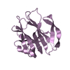
- PDB-3aly: Crystal Structure of RNase HI from Sulfolobus tokodaii with C-ter... -
+ Open data

- Basic information
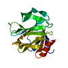
| Entry | Database: PDB / ID: 3aly | ||||||
|---|---|---|---|---|---|---|---|
| Title | Crystal Structure of RNase HI from Sulfolobus tokodaii with C-terminal deletion | ||||||
 Components Components | Putative uncharacterized protein ST0753 | ||||||
 Keywords Keywords | HYDROLASE / thermostable RNase HI / C-terminal anchor deletion | ||||||
| Function / homology |  Function and homology information Function and homology informationribonuclease H / RNA-DNA hybrid ribonuclease activity / DNA binding / RNA binding / metal ion binding / cytoplasm Similarity search - Function | ||||||
| Biological species |   Sulfolobus tokodaii (archaea) Sulfolobus tokodaii (archaea) | ||||||
| Method |  X-RAY DIFFRACTION / SYNCHROTRON / MOLECULAR REPLACEMENT / Resolution: 1.66 Å X-RAY DIFFRACTION / SYNCHROTRON / MOLECULAR REPLACEMENT / Resolution: 1.66 Å | ||||||
 Authors Authors | Angkawidjaja, C. / Takano, K. / Kanaya, S. | ||||||
 Citation Citation | Journal: Plos One / Year: 2011 Title: Stabilization by fusion to the C-terminus of hyperthermophile Sulfolobus tokodaii RNase HI: a possibility of protein stabilization tag Authors: Takano, K. / Okamoto, T. / Okada, J. / Tanaka, S. / Angkawidjaja, C. / Koga, Y. / Kanaya, S. | ||||||
| History |
|
- Structure visualization
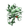
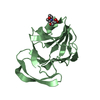
| Structure viewer | Molecule: MolmilJmol/JSmol |
|---|
- Downloads & links
-Download
| PDBx/mmCIF format | 3aly.cif.gz | 72.4 KB | Display | PDBx/mmCIF format |
|---|---|---|---|---|
| PDB format | pdb3aly.ent.gz | 53.7 KB | Display | PDB format |
| PDBx/mmJSON format | 3aly.json.gz | Tree view | PDBx/mmJSON format | |
| Others |  Other downloads Other downloads |
-Validation report
| Arichive directory | https://data.pdbj.org/pub/pdb/validation_reports/al/3alyftp://data.pdbj.org/pub/pdb/validation_reports/al/3aly | HTTPS FTP |
|---|
-Related structure data
| Related structure data | 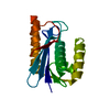 2ehgS S: Starting model for refinement |
|---|---|
| Similar structure data |


-Links
 PDBj
PDBj
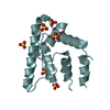
- Assembly
| Deposited unit | 
| ||||||||
|---|---|---|---|---|---|---|---|---|---|
| 1 | 
| ||||||||
| 2 | 
| ||||||||
| 3 |
| ||||||||
| Unit cell |
|
-Components
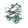
| #1: Protein | Mass: 16235.057 Da / Num. of mol.: 2 / Fragment: residues 1-143 Source method: isolated from a genetically manipulated source Source: (gene. exp.) Sulfolobus tokodaii (archaea) / Strain: str. 7 / Gene: ST0753 / Plasmid: pET25b+ / Production host:  References: UniProt: Q973Z8, UniProt: F9VN79*PLUS, ribonuclease H #2: Water | ChemComp-HOH / |  Mass: 18.015 Da / Num. of mol.: 254 / Source method: isolated from a natural source / Formula: H2O Mass: 18.015 Da / Num. of mol.: 254 / Source method: isolated from a natural source / Formula: H2O |
|---|
-Experimental details
-Experiment
| Experiment | Method: X-RAY DIFFRACTION / Number of used crystals: 1 |
|---|
- Sample preparation
| Crystal | Density Matthews: 2.25 Å3/Da / Density % sol: 45.33 % |
|---|---|
| Crystal grow | Temperature: 277 K / Method: vapor diffusion, sitting drop / pH: 5.5 Details: 20% PEG 3000, 0.1M citrate, pH 5.5, VAPOR DIFFUSION, SITTING DROP, temperature 277K |
-Data collection
| Diffraction | Mean temperature: 100 K |
|---|---|
| Diffraction source | Source: SYNCHROTRON / Site: SPring-8  / Beamline: BL38B1 / Wavelength: 1 Å / Beamline: BL38B1 / Wavelength: 1 Å |
| Detector | Type: ADSC QUANTUM 210 / Detector: CCD / Date: Dec 4, 2009 / Details: mirrors |
| Radiation | Monochromator: mirrors / Protocol: SINGLE WAVELENGTH / Monochromatic (M) / Laue (L): M / Scattering type: x-ray |
| Radiation wavelength | Wavelength: 1 Å / Relative weight: 1 |
| Reflection | Resolution: 1.66→50 Å / Num. obs: 32375 / % possible obs: 96.2 % / Observed criterion σ(F): 5 / Observed criterion σ(I): 5 / Redundancy: 3.9 % / Rmerge(I) obs: 0.034 / Rsym value: 0.031 / Net I/σ(I): 37.21 |
| Reflection shell | Resolution: 1.66→1.72 Å / Redundancy: 3.9 % / Rmerge(I) obs: 0.218 / Mean I/σ(I) obs: 6.14 / Num. unique all: 3175 / Rsym value: 0.193 / % possible all: 94.3 |
- Processing
| Software |
| |||||||||||||||||||||||||||||||||||||||||||||||||||||||||||||||||
|---|---|---|---|---|---|---|---|---|---|---|---|---|---|---|---|---|---|---|---|---|---|---|---|---|---|---|---|---|---|---|---|---|---|---|---|---|---|---|---|---|---|---|---|---|---|---|---|---|---|---|---|---|---|---|---|---|---|---|---|---|---|---|---|---|---|---|
| Refinement | Method to determine structure: MOLECULAR REPLACEMENT Starting model: 2EHG Resolution: 1.66→20.48 Å / Cor.coef. Fo:Fc: 0.951 / Cor.coef. Fo:Fc free: 0.926 / SU B: 2.076 / SU ML: 0.073 / Cross valid method: THROUGHOUT / ESU R Free: 0.122 Stereochemistry target values: MAXIMUM LIKELIHOOD WITH PHASES Details: HYDROGENS HAVE BEEN ADDED IN THE RIDING POSITIONS
| |||||||||||||||||||||||||||||||||||||||||||||||||||||||||||||||||
| Solvent computation | Ion probe radii: 0.8 Å / Shrinkage radii: 0.8 Å / VDW probe radii: 1.4 Å / Solvent model: MASK | |||||||||||||||||||||||||||||||||||||||||||||||||||||||||||||||||
| Displacement parameters | Biso mean: 25.003 Å2
| |||||||||||||||||||||||||||||||||||||||||||||||||||||||||||||||||
| Refinement step | Cycle: LAST / Resolution: 1.66→20.48 Å
| |||||||||||||||||||||||||||||||||||||||||||||||||||||||||||||||||
| Refine LS restraints |
| |||||||||||||||||||||||||||||||||||||||||||||||||||||||||||||||||
| LS refinement shell | Resolution: 1.657→1.7 Å / Total num. of bins used: 20
|
