 Movie
Movie Controller

[English] 日本語
 Yorodumi
- PDB-7ec8: Polyethylene terephthalate hydrolyzing lipase PET2 mutant - F105R... -
+ Open data

- Basic information
| Entry | Database: PDB / ID: 7ec8 | ||||||
|---|---|---|---|---|---|---|---|
| Title | Polyethylene terephthalate hydrolyzing lipase PET2 mutant - F105R-E110K | ||||||
 Components Components | LipIAF5-2 | ||||||
 Keywords Keywords | HYDROLASE / Polyethylene terephthalate hydrolase / lipase | ||||||
| Function / homology | Cutinase / PET hydrolase-like / : / carboxylic ester hydrolase activity / Alpha/Beta hydrolase fold / LipIAF5-2 Function and homology information Function and homology information | ||||||
| Biological species |  uncultured bacterium (environmental samples) uncultured bacterium (environmental samples) | ||||||
| Method |  X-RAY DIFFRACTION / SYNCHROTRON / MOLECULAR REPLACEMENT / Resolution: 1.35 Å X-RAY DIFFRACTION / SYNCHROTRON / MOLECULAR REPLACEMENT / Resolution: 1.35 Å | ||||||
 Authors Authors | Nakamura, A. | ||||||
 Citation Citation | Journal: Acs Catalysis / Year: 2021 Title: Positive Charge Introduction on the Surface of Thermostabilized PET Hydrolase Facilitates PET Binding and Degradation. Authors: Nakamura, A. | ||||||
| History |
|
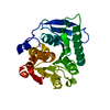
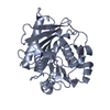
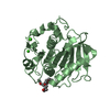
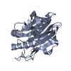
- Structure visualization
| Structure viewer | Molecule: MolmilJmol/JSmol |
|---|
- Downloads & links
-Download
| PDBx/mmCIF format | 7ec8.cif.gz | 135.7 KB | Display | PDBx/mmCIF format |
|---|---|---|---|---|
| PDB format | pdb7ec8.ent.gz | 102.6 KB | Display | PDB format |
| PDBx/mmJSON format | 7ec8.json.gz | Tree view | PDBx/mmJSON format | |
| Others |  Other downloads Other downloads |
-Validation report
| Arichive directory | https://data.pdbj.org/pub/pdb/validation_reports/ec/7ec8ftp://data.pdbj.org/pub/pdb/validation_reports/ec/7ec8 | HTTPS FTP |
|---|
-Related structure data


-Links
 PDBj
PDBj- Assembly
| Deposited unit | 
| ||||||||||||
|---|---|---|---|---|---|---|---|---|---|---|---|---|---|
| 1 | 
| ||||||||||||
| 2 | 
| ||||||||||||
| Unit cell |
|
-Components
| #1: Protein | Mass: 31610.246 Da / Num. of mol.: 2 / Mutation: F105R, E110K Source method: isolated from a genetically manipulated source Source: (gene. exp.) uncultured bacterium (environmental samples)Gene: lipIAF5-2 / Plasmid: pET27b / Production host: #2: Chemical | ChemComp-EDO /   Mass: 62.068 Da / Num. of mol.: 14 / Source method: obtained synthetically / Formula: C2H6O2 Mass: 62.068 Da / Num. of mol.: 14 / Source method: obtained synthetically / Formula: C2H6O2#3: Chemical |   Mass: 96.063 Da / Num. of mol.: 3 / Source method: obtained synthetically / Formula: SO4 Mass: 96.063 Da / Num. of mol.: 3 / Source method: obtained synthetically / Formula: SO4#4: Water | ChemComp-HOH / |  Mass: 18.015 Da / Num. of mol.: 655 / Source method: isolated from a natural source / Formula: H2O Mass: 18.015 Da / Num. of mol.: 655 / Source method: isolated from a natural source / Formula: H2OHas ligand of interest | N | Has protein modification | Y | |
|---|
-Experimental details
-Experiment
| Experiment | Method: X-RAY DIFFRACTION / Number of used crystals: 1 |
|---|
- Sample preparation
| Crystal | Density Matthews: 2 Å3/Da / Density % sol: 38.44 % |
|---|---|
| Crystal grow | Temperature: 293 K / Method: vapor diffusion, sitting drop / pH: 8 / Details: 1 M ammonium sulfate in 100 mM Tris-HCl pH 8.0 |
-Data collection
| Diffraction | Mean temperature: 100 K / Serial crystal experiment: N |
|---|---|
| Diffraction source | Source: SYNCHROTRON / Site: AichiSR  / Beamline: BL2S1 / Wavelength: 1.12 Å / Beamline: BL2S1 / Wavelength: 1.12 Å |
| Detector | Type: ADSC QUANTUM 270 / Detector: CCD / Date: Oct 14, 2020 |
| Radiation | Protocol: SINGLE WAVELENGTH / Monochromatic (M) / Laue (L): M / Scattering type: x-ray |
| Radiation wavelength | Wavelength: 1.12 Å / Relative weight: 1 |
| Reflection | Resolution: 1.35→33.4 Å / Num. obs: 111811 / % possible obs: 99.5 % / Redundancy: 13.9 % / Biso Wilson estimate: 13.19 Å2 / CC1/2: 1 / Rmerge(I) obs: 0.077 / Net I/σ(I): 23.9 |
| Reflection shell | Resolution: 1.35→1.43 Å / Redundancy: 13.6 % / Rmerge(I) obs: 1.24 / Mean I/σ(I) obs: 2.3 / Num. unique obs: 17635 / CC1/2: 0.772 / % possible all: 97.9 |
- Processing
| Software |
| |||||||||||||||||||||||||||||||||||||||||||||||||||||||||||||||||||||||||||||||||||||||||||||||||||||||||||||||||||||||||||||||||||||||||||||||||||||||||||||||||||||||||||||||||||||||||||||||||||||||||||||||||||||||||
|---|---|---|---|---|---|---|---|---|---|---|---|---|---|---|---|---|---|---|---|---|---|---|---|---|---|---|---|---|---|---|---|---|---|---|---|---|---|---|---|---|---|---|---|---|---|---|---|---|---|---|---|---|---|---|---|---|---|---|---|---|---|---|---|---|---|---|---|---|---|---|---|---|---|---|---|---|---|---|---|---|---|---|---|---|---|---|---|---|---|---|---|---|---|---|---|---|---|---|---|---|---|---|---|---|---|---|---|---|---|---|---|---|---|---|---|---|---|---|---|---|---|---|---|---|---|---|---|---|---|---|---|---|---|---|---|---|---|---|---|---|---|---|---|---|---|---|---|---|---|---|---|---|---|---|---|---|---|---|---|---|---|---|---|---|---|---|---|---|---|---|---|---|---|---|---|---|---|---|---|---|---|---|---|---|---|---|---|---|---|---|---|---|---|---|---|---|---|---|---|---|---|---|---|---|---|---|---|---|---|---|---|---|---|---|---|---|---|---|
| Refinement | Method to determine structure: MOLECULAR REPLACEMENT / Resolution: 1.35→33.4 Å / SU ML: 0.1685 / Cross valid method: FREE R-VALUE / σ(F): 1.36 / Phase error: 20.3612 / Stereochemistry target values: GeoStd + Monomer Library
| |||||||||||||||||||||||||||||||||||||||||||||||||||||||||||||||||||||||||||||||||||||||||||||||||||||||||||||||||||||||||||||||||||||||||||||||||||||||||||||||||||||||||||||||||||||||||||||||||||||||||||||||||||||||||
| Solvent computation | Shrinkage radii: 0.9 Å / VDW probe radii: 1.11 Å / Solvent model: FLAT BULK SOLVENT MODEL | |||||||||||||||||||||||||||||||||||||||||||||||||||||||||||||||||||||||||||||||||||||||||||||||||||||||||||||||||||||||||||||||||||||||||||||||||||||||||||||||||||||||||||||||||||||||||||||||||||||||||||||||||||||||||
| Displacement parameters | Biso mean: 17.56 Å2 | |||||||||||||||||||||||||||||||||||||||||||||||||||||||||||||||||||||||||||||||||||||||||||||||||||||||||||||||||||||||||||||||||||||||||||||||||||||||||||||||||||||||||||||||||||||||||||||||||||||||||||||||||||||||||
| Refinement step | Cycle: LAST / Resolution: 1.35→33.4 Å
| |||||||||||||||||||||||||||||||||||||||||||||||||||||||||||||||||||||||||||||||||||||||||||||||||||||||||||||||||||||||||||||||||||||||||||||||||||||||||||||||||||||||||||||||||||||||||||||||||||||||||||||||||||||||||
| Refine LS restraints |
| |||||||||||||||||||||||||||||||||||||||||||||||||||||||||||||||||||||||||||||||||||||||||||||||||||||||||||||||||||||||||||||||||||||||||||||||||||||||||||||||||||||||||||||||||||||||||||||||||||||||||||||||||||||||||
| LS refinement shell |
|
