 Movie
Movie Controller

[English] 日本語
 Yorodumi
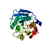
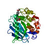
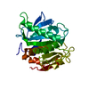
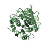
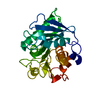
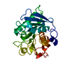
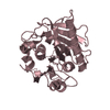
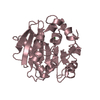
- PDB-5yns: Crystal structure of PETase R280A mutant from Ideonella sakaiensis -
+ Open data

- Basic information
| Entry | Database: PDB / ID: 5yns | ||||||
|---|---|---|---|---|---|---|---|
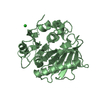
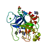
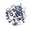
| Title | Crystal structure of PETase R280A mutant from Ideonella sakaiensis | ||||||
 Components Components | Poly(ethylene terephthalate) hydrolase | ||||||
 Keywords Keywords | HYDROLASE | ||||||
| Function / homology |  Function and homology information Function and homology informationacetylesterase activity / poly(ethylene terephthalate) hydrolase / carboxylic ester hydrolase activity / xenobiotic catabolic process / extracellular region Similarity search - Function | ||||||
| Biological species |  Ideonella sakaiensis (bacteria) Ideonella sakaiensis (bacteria) | ||||||
| Method |  X-RAY DIFFRACTION / SYNCHROTRON / MOLECULAR REPLACEMENT / Resolution: 1.36 Å X-RAY DIFFRACTION / SYNCHROTRON / MOLECULAR REPLACEMENT / Resolution: 1.36 Å | ||||||
 Authors Authors | Joo, S. / Kim, K.-J. | ||||||
 Citation Citation | Journal: Nat Commun / Year: 2018 Title: Structural insight into molecular mechanism of poly(ethylene terephthalate) degradation. Authors: Joo, S. / Cho, I.J. / Seo, H. / Son, H.F. / Sagong, H.-Y. / Shin, T.J. / Choi, S.Y. / Lee, S.Y. / Kim, K.-J. | ||||||
| History |
|
- Structure visualization
| Structure viewer | Molecule: MolmilJmol/JSmol |
|---|
- Downloads & links
-Download
| PDBx/mmCIF format | 5yns.cif.gz | 69.8 KB | Display | PDBx/mmCIF format |
|---|---|---|---|---|
| PDB format | pdb5yns.ent.gz | 49.3 KB | Display | PDB format |
| PDBx/mmJSON format | 5yns.json.gz | Tree view | PDBx/mmJSON format | |
| Others |  Other downloads Other downloads |
-Validation report
| Arichive directory | https://data.pdbj.org/pub/pdb/validation_reports/yn/5ynsftp://data.pdbj.org/pub/pdb/validation_reports/yn/5yns | HTTPS FTP |
|---|
-Related structure data
| Related structure data |  5xjhSC S: Starting model for refinement C: citing same article ( |
|---|---|
| Similar structure data |


-Links
 PDBj
PDBj
- Assembly
| Deposited unit | 
| ||||||||
|---|---|---|---|---|---|---|---|---|---|
| 1 |
| ||||||||
| Unit cell |
|
-Components
| #1: Protein | Mass: 27590.596 Da / Num. of mol.: 1 / Fragment: a/b hydrolase superfamily / Mutation: R280A Source method: isolated from a genetically manipulated source Source: (gene. exp.) Ideonella sakaiensis (strain 201-F6) (bacteria)Strain: 201-F6 / Gene: ISF6_4831 / Plasmid: pET15b / Production host: References: UniProt: A0A0K8P6T7, poly(ethylene terephthalate) hydrolase |
|---|---|
| #2: Water | ChemComp-HOH /  Mass: 18.015 Da / Num. of mol.: 294 / Source method: isolated from a natural source / Formula: H2O Mass: 18.015 Da / Num. of mol.: 294 / Source method: isolated from a natural source / Formula: H2O |
| Has protein modification | Y |
-Experimental details
-Experiment
| Experiment | Method: X-RAY DIFFRACTION / Number of used crystals: 1 |
|---|
- Sample preparation
| Crystal | Density Matthews: 2.59 Å3/Da / Density % sol: 52.51 % |
|---|---|
| Crystal grow | Temperature: 293 K / Method: vapor diffusion, hanging drop / pH: 5.5 / Details: BIS-Tris, Ammonium acetate, PEG 10000 |
-Data collection
| Diffraction | Mean temperature: 100 K |
|---|---|
| Diffraction source | Source: SYNCHROTRON / Site: PAL/PLS  / Beamline: 7A (6B, 6C1) / Wavelength: 0.97934 Å / Beamline: 7A (6B, 6C1) / Wavelength: 0.97934 Å |
| Detector | Type: ADSC QUANTUM 270 / Detector: CCD / Date: Oct 24, 2017 |
| Radiation | Protocol: SINGLE WAVELENGTH / Monochromatic (M) / Laue (L): M / Scattering type: x-ray |
| Radiation wavelength | Wavelength: 0.97934 Å / Relative weight: 1 |
| Reflection | Resolution: 1.36→50 Å / Num. obs: 62494 / % possible obs: 98.7 % / Redundancy: 9.3 % / Rmerge(I) obs: 0.064 / Net I/σ(I): 40.79 |
| Reflection shell | Rmerge(I) obs: 0.374 |
- Processing
| Software |
| ||||||||||||||||||||||||||||||||||||||||||||||||||||||||||||
|---|---|---|---|---|---|---|---|---|---|---|---|---|---|---|---|---|---|---|---|---|---|---|---|---|---|---|---|---|---|---|---|---|---|---|---|---|---|---|---|---|---|---|---|---|---|---|---|---|---|---|---|---|---|---|---|---|---|---|---|---|---|
| Refinement | Method to determine structure: MOLECULAR REPLACEMENT Starting model: 5XJH Resolution: 1.36→27.3 Å / Cor.coef. Fo:Fc: 0.971 / Cor.coef. Fo:Fc free: 0.96 / SU B: 0.858 / SU ML: 0.034 / Cross valid method: THROUGHOUT / σ(F): 0 / ESU R: 0.048 / ESU R Free: 0.052 Details: HYDROGENS HAVE BEEN ADDED IN THE RIDING POSITIONS U VALUES : REFINED INDIVIDUALLY
| ||||||||||||||||||||||||||||||||||||||||||||||||||||||||||||
| Solvent computation | Ion probe radii: 0.8 Å / Shrinkage radii: 0.8 Å / VDW probe radii: 1.2 Å | ||||||||||||||||||||||||||||||||||||||||||||||||||||||||||||
| Displacement parameters | Biso max: 58.25 Å2 / Biso mean: 14.344 Å2 / Biso min: 2 Å2
| ||||||||||||||||||||||||||||||||||||||||||||||||||||||||||||
| Refinement step | Cycle: final / Resolution: 1.36→27.3 Å
| ||||||||||||||||||||||||||||||||||||||||||||||||||||||||||||
| Refine LS restraints |
| ||||||||||||||||||||||||||||||||||||||||||||||||||||||||||||
| LS refinement shell | Resolution: 1.36→1.396 Å / Rfactor Rfree error: 0 / Total num. of bins used: 20
|
