 Movie
Movie Controller

[English] 日本語
 Yorodumi
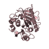
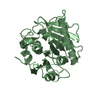
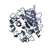
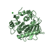
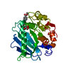
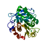
- PDB-5xg0: Crystal structure of a novel PET hydrolase from Ideonella sakaien... -
+ Open data

- Basic information
| Entry | Database: PDB / ID: 5xg0 | ||||||
|---|---|---|---|---|---|---|---|
| Title | Crystal structure of a novel PET hydrolase from Ideonella sakaiensis 201-F6 | ||||||
 Components Components | Poly(ethylene terephthalate) hydrolase | ||||||
 Keywords Keywords | HYDROLASE / metal-binding / substrate binding / acidocalcisomal pyrophosphatase / inhibitor | ||||||
| Function / homology |  Function and homology information Function and homology informationacetylesterase activity / poly(ethylene terephthalate) hydrolase / carboxylic ester hydrolase activity / xenobiotic catabolic process / extracellular region Similarity search - Function | ||||||
| Biological species |  Ideonella sakaiensis (bacteria) Ideonella sakaiensis (bacteria) | ||||||
| Method |  X-RAY DIFFRACTION / SYNCHROTRON / MOLECULAR REPLACEMENT / molecular replacement / Resolution: 1.58 Å X-RAY DIFFRACTION / SYNCHROTRON / MOLECULAR REPLACEMENT / molecular replacement / Resolution: 1.58 Å | ||||||
 Authors Authors | Han, X. / Liu, W.D. / Zheng, Y.Y. / Chen, C.C. / Guo, R.T. | ||||||
 Citation Citation | Journal: Nat Commun / Year: 2017 Title: Structural insight into catalytic mechanism of PET hydrolase Authors: Han, X. / Liu, W. / Huang, J.W. / Ma, J. / Zheng, Y. / Ko, T.P. / Xu, L. / Cheng, Y.S. / Chen, C.C. / Guo, R.T. | ||||||
| History |
|
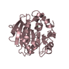
- Structure visualization
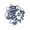
| Structure viewer | Molecule: MolmilJmol/JSmol |
|---|
- Downloads & links
-Download
| PDBx/mmCIF format | 5xg0.cif.gz | 179.3 KB | Display | PDBx/mmCIF format |
|---|---|---|---|---|
| PDB format | pdb5xg0.ent.gz | 139.3 KB | Display | PDB format |
| PDBx/mmJSON format | 5xg0.json.gz | Tree view | PDBx/mmJSON format | |
| Others |  Other downloads Other downloads |
-Validation report
| Arichive directory | https://data.pdbj.org/pub/pdb/validation_reports/xg/5xg0ftp://data.pdbj.org/pub/pdb/validation_reports/xg/5xg0 | HTTPS FTP |
|---|
-Related structure data
| Related structure data |  5xfyC  5xfzC  5xh2C  5xh3C  4wfiS  5xfx C: citing same article ( S: Starting model for refinement |
|---|---|
| Similar structure data |


-Links
 PDBj
PDBj
- Assembly
| Deposited unit | 
| ||||||||
|---|---|---|---|---|---|---|---|---|---|
| 1 | 
| ||||||||
| 2 | 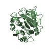
| ||||||||
| 3 | 
| ||||||||
| Unit cell |
|
-Components
| #1: Protein | Mass: 27866.930 Da / Num. of mol.: 3 / Fragment: UNP residues 30-290 Source method: isolated from a genetically manipulated source Source: (gene. exp.) Ideonella sakaiensis (strain 201-F6) (bacteria)Strain: 201-F6 / Gene: ISF6_4831 / Plasmid: pET32a / Production host: References: UniProt: A0A0K8P6T7, poly(ethylene terephthalate) hydrolase #2: Water | ChemComp-HOH / |  Mass: 18.015 Da / Num. of mol.: 1124 / Source method: isolated from a natural source / Formula: H2O Mass: 18.015 Da / Num. of mol.: 1124 / Source method: isolated from a natural source / Formula: H2OHas protein modification | Y | |
|---|
-Experimental details
-Experiment
| Experiment | Method: X-RAY DIFFRACTION / Number of used crystals: 1 |
|---|
- Sample preparation
| Crystal | Density Matthews: 2.01 Å3/Da / Density % sol: 38.83 % / Mosaicity: 0.29 ° |
|---|---|
| Crystal grow | Temperature: 298 K / Method: vapor diffusion, sitting drop / pH: 6 / Details: Polyethylene Glycol 6000, Glycerol, MES |
-Data collection
| Diffraction | Mean temperature: 100 K |
|---|---|
| Diffraction source | Source: SYNCHROTRON / Site: NSRRC  / Beamline: BL15A1 / Wavelength: 1 Å / Beamline: BL15A1 / Wavelength: 1 Å |
| Detector | Type: RAYONIX MX-300 / Detector: CCD / Date: Oct 29, 2016 |
| Radiation | Monochromator: GRAPHITE / Protocol: SINGLE WAVELENGTH / Monochromatic (M) / Laue (L): M / Scattering type: x-ray |
| Radiation wavelength | Wavelength: 1 Å / Relative weight: 1 |
| Reflection | Resolution: 1.58→25 Å / Num. obs: 92787 / % possible obs: 99.8 % / Redundancy: 5.4 % / Rmerge(I) obs: 0.039 / Rpim(I) all: 0.018 / Rrim(I) all: 0.043 / Χ2: 1.54 / Net I/σ(I): 32.2 / Num. measured all: 499698 |
| Reflection shell | Resolution: 1.58→1.64 Å / Redundancy: 5.4 % / Rmerge(I) obs: 0.076 / Num. unique obs: 9169 / Rpim(I) all: 0.036 / Rrim(I) all: 0.084 / Χ2: 1.74 / % possible all: 100 |
-Phasing
| Phasing | Method: molecular replacement |
|---|
- Processing
| Software |
| ||||||||||||||||||||||||||||||||||||||||||||||||||||||||||||
|---|---|---|---|---|---|---|---|---|---|---|---|---|---|---|---|---|---|---|---|---|---|---|---|---|---|---|---|---|---|---|---|---|---|---|---|---|---|---|---|---|---|---|---|---|---|---|---|---|---|---|---|---|---|---|---|---|---|---|---|---|---|
| Refinement | Method to determine structure: MOLECULAR REPLACEMENT Starting model: 4WFI Resolution: 1.58→25 Å / Cor.coef. Fo:Fc: 0.969 / Cor.coef. Fo:Fc free: 0.961 / SU B: 1.085 / SU ML: 0.04 / Cross valid method: THROUGHOUT / σ(F): 0 / ESU R: 0.075 / ESU R Free: 0.073 Details: HYDROGENS HAVE BEEN ADDED IN THE RIDING POSITIONS U VALUES : REFINED INDIVIDUALLY
| ||||||||||||||||||||||||||||||||||||||||||||||||||||||||||||
| Solvent computation | Ion probe radii: 0.8 Å / Shrinkage radii: 0.8 Å / VDW probe radii: 1.2 Å | ||||||||||||||||||||||||||||||||||||||||||||||||||||||||||||
| Displacement parameters | Biso max: 81.08 Å2 / Biso mean: 11.229 Å2 / Biso min: 1 Å2
| ||||||||||||||||||||||||||||||||||||||||||||||||||||||||||||
| Refinement step | Cycle: final / Resolution: 1.58→25 Å
| ||||||||||||||||||||||||||||||||||||||||||||||||||||||||||||
| Refine LS restraints |
| ||||||||||||||||||||||||||||||||||||||||||||||||||||||||||||
| LS refinement shell | Resolution: 1.58→1.621 Å / Rfactor Rfree error: 0 / Total num. of bins used: 20
|
