 Movie
Movie Controller


+ Open data

- Basic information
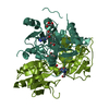
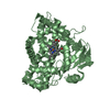
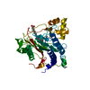
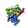
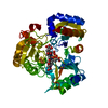
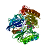
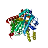
| Entry | Database: PDB / ID: 7ebl | ||||||
|---|---|---|---|---|---|---|---|
| Title | Bacterial STING in complex with c-di-GMP | ||||||
 Components Components | STING | ||||||
 Keywords Keywords | SIGNALING PROTEIN / second messenger / complex / cyclic dinucleotide / antiviral pathway | ||||||
| Function / homology | Chem-C2E Function and homology information Function and homology information | ||||||
| Biological species |  Myroides sp. ZB35 (bacteria) Myroides sp. ZB35 (bacteria) | ||||||
| Method |  X-RAY DIFFRACTION / SYNCHROTRON / MOLECULAR REPLACEMENT / Resolution: 2.17 Å X-RAY DIFFRACTION / SYNCHROTRON / MOLECULAR REPLACEMENT / Resolution: 2.17 Å | ||||||
 Authors Authors | Ko, T.-P. / Wang, Y.-C. / Yang, C.-S. / Hou, M.-H. / Chen, Y. | ||||||
| Funding support |  Taiwan, 1items Taiwan, 1items
| ||||||
 Citation Citation | Journal: Nat Commun / Year: 2022 Title: Crystal structure and functional implication of bacterial STING. Authors: Ko, T.-P. / Wang, Y.-C. / Yang, C.-S. / Hou, M.-H. / Chen, C.-J. / Chiu, Y.-F. / Chen, Y. | ||||||
| History |
|
- Structure visualization
| Structure viewer | Molecule: MolmilJmol/JSmol |
|---|
- Downloads & links
-Download
| PDBx/mmCIF format | 7ebl.cif.gz | 105.9 KB | Display | PDBx/mmCIF format |
|---|---|---|---|---|
| PDB format | pdb7ebl.ent.gz | 64.1 KB | Display | PDB format |
| PDBx/mmJSON format | 7ebl.json.gz | Tree view | PDBx/mmJSON format | |
| Others |  Other downloads Other downloads |
-Validation report
| Arichive directory | https://data.pdbj.org/pub/pdb/validation_reports/eb/7eblftp://data.pdbj.org/pub/pdb/validation_reports/eb/7ebl | HTTPS FTP |
|---|
-Related structure data
| Related structure data |  7ebdSC S: Starting model for refinement C: citing same article ( |
|---|---|
| Similar structure data |


-Links
 PDBj
PDBj
- Assembly
| Deposited unit | 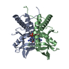
| ||||||||||||||||||||||||||||||||||||||||||||||||||||||||||
|---|---|---|---|---|---|---|---|---|---|---|---|---|---|---|---|---|---|---|---|---|---|---|---|---|---|---|---|---|---|---|---|---|---|---|---|---|---|---|---|---|---|---|---|---|---|---|---|---|---|---|---|---|---|---|---|---|---|---|---|
| 1 |
| ||||||||||||||||||||||||||||||||||||||||||||||||||||||||||
| Unit cell |
| ||||||||||||||||||||||||||||||||||||||||||||||||||||||||||
| Noncrystallographic symmetry (NCS) | NCS domain:
NCS domain segments:
NCS oper: (Code: givenMatrix: (-0.9995781002, -0.0166128385123, -0.0238250959639), (0.0200411056817, -0.988214815931, -0.151755829072), (-0.0210232177408, -0.152169284584, 0.98813082795)Vector: 0. ...NCS oper: (Code: given Matrix: (-0.9995781002, -0.0166128385123, -0.0238250959639), Vector: |
-Components
| #1: Protein | Mass: 19183.301 Da / Num. of mol.: 2 Source method: isolated from a genetically manipulated source Source: (gene. exp.) Myroides sp. ZB35 (bacteria)Production host: #2: Chemical | ChemComp-C2E / | 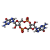  Mass: 690.411 Da / Num. of mol.: 1 / Source method: obtained synthetically / Formula: C20H24N10O14P2 / Feature type: SUBJECT OF INVESTIGATION Mass: 690.411 Da / Num. of mol.: 1 / Source method: obtained synthetically / Formula: C20H24N10O14P2 / Feature type: SUBJECT OF INVESTIGATION#3: Water | ChemComp-HOH / |  Mass: 18.015 Da / Num. of mol.: 228 / Source method: isolated from a natural source / Formula: H2O Mass: 18.015 Da / Num. of mol.: 228 / Source method: isolated from a natural source / Formula: H2OHas ligand of interest | Y | Sequence details | The sequence has been deposited to NCBI under accession number WP_071307203. | |
|---|
-Experimental details
-Experiment
| Experiment | Method: X-RAY DIFFRACTION / Number of used crystals: 1 |
|---|
- Sample preparation
| Crystal | Density Matthews: 2.18 Å3/Da / Density % sol: 43.57 % |
|---|---|
| Crystal grow | Temperature: 277 K / Method: vapor diffusion, sitting drop Details: 0.1M sodium cacodylate trihydrate pH 6.5, 0.2M sodium acetate trihydrate, 15% w/v polyethylene glycol 8000 |
-Data collection
| Diffraction | Mean temperature: 100 K / Serial crystal experiment: N |
|---|---|
| Diffraction source | Source: SYNCHROTRON / Site: NSRRC / Beamline: TPS 05A / Wavelength: 0.9998 Å |
| Detector | Type: RAYONIX MX300-HS / Detector: CCD / Date: Dec 10, 2020 |
| Radiation | Protocol: SINGLE WAVELENGTH / Monochromatic (M) / Laue (L): M / Scattering type: x-ray |
| Radiation wavelength | Wavelength: 0.9998 Å / Relative weight: 1 |
| Reflection | Resolution: 2.17→30 Å / Num. obs: 17092 / % possible obs: 98.3 % / Redundancy: 3.5 % / Biso Wilson estimate: 29.9 Å2 / Rmerge(I) obs: 0.082 / Rpim(I) all: 0.052 / Net I/σ(I): 15.7 |
| Reflection shell | Resolution: 2.17→2.25 Å / Redundancy: 3 % / Rmerge(I) obs: 0.32 / Mean I/σ(I) obs: 3.1 / Num. unique obs: 1680 / CC1/2: 0.859 / Rpim(I) all: 0.215 / % possible all: 96.9 |
- Processing
| Software |
| |||||||||||||||||||||||||||||||||||||||||||||||||
|---|---|---|---|---|---|---|---|---|---|---|---|---|---|---|---|---|---|---|---|---|---|---|---|---|---|---|---|---|---|---|---|---|---|---|---|---|---|---|---|---|---|---|---|---|---|---|---|---|---|---|
| Refinement | Method to determine structure: MOLECULAR REPLACEMENT Starting model: 7EBD Resolution: 2.17→24.18 Å / SU ML: 0.2156 / Cross valid method: FREE R-VALUE / σ(F): 1.4 / Phase error: 23.0571 Stereochemistry target values: GeoStd + Monomer Library + CDL v1.2
| |||||||||||||||||||||||||||||||||||||||||||||||||
| Solvent computation | Shrinkage radii: 0.9 Å / VDW probe radii: 1.11 Å / Solvent model: FLAT BULK SOLVENT MODEL | |||||||||||||||||||||||||||||||||||||||||||||||||
| Displacement parameters | Biso mean: 34.75 Å2 | |||||||||||||||||||||||||||||||||||||||||||||||||
| Refinement step | Cycle: LAST / Resolution: 2.17→24.18 Å
| |||||||||||||||||||||||||||||||||||||||||||||||||
| Refine LS restraints |
| |||||||||||||||||||||||||||||||||||||||||||||||||
| Refine LS restraints NCS | Type: Torsion NCS / Rms dev position: 1.15037369713 Å | |||||||||||||||||||||||||||||||||||||||||||||||||
| LS refinement shell |
|
