 Movie
Movie Controller

[English] 日本語
 Yorodumi
- PDB-7ebi: Chitin-specific solute binding protein from Vibrio harveyi co-cry... -
+ Open data

- Basic information
| Entry | Database: PDB / ID: 7ebi | |||||||||
|---|---|---|---|---|---|---|---|---|---|---|
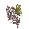
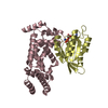
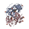
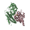
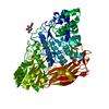
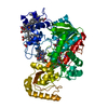
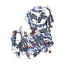
| Title | Chitin-specific solute binding protein from Vibrio harveyi co-crystalized with chitotetraose. | |||||||||
 Components Components | Peptide ABC transporter, periplasmic peptide-binding protein | |||||||||
 Keywords Keywords | SUGAR BINDING PROTEIN / complex / chitin / periplasmic solute-binding protein / Vibrios | |||||||||
| Function / homology |  Function and homology information Function and homology informationATP-binding cassette (ABC) transporter complex / transmembrane transport / outer membrane-bounded periplasmic space Similarity search - Function | |||||||||
| Biological species |  Vibrio harveyi (bacteria) Vibrio harveyi (bacteria) | |||||||||
| Method |  X-RAY DIFFRACTION / SYNCHROTRON / MOLECULAR REPLACEMENT / Resolution: 1.5 Å X-RAY DIFFRACTION / SYNCHROTRON / MOLECULAR REPLACEMENT / Resolution: 1.5 Å | |||||||||
 Authors Authors | Kitaoku, Y. / Ubonbal, P. / Tran, L.T. / Robinson, R.C. / Suginta, W. | |||||||||
| Funding support |  Thailand, 2items Thailand, 2items
| |||||||||
 Citation Citation | Journal: J.Biol.Chem. / Year: 2021 Title: A structural model for (GlcNAc) 2 translocation via a periplasmic chitooligosaccharide-binding protein from marine Vibrio bacteria. Authors: Kitaoku, Y. / Fukamizo, T. / Kumsaoad, S. / Ubonbal, P. / Robinson, R.C. / Suginta, W. | |||||||||
| History |
|
- Structure visualization
| Structure viewer | Molecule: MolmilJmol/JSmol |
|---|
- Downloads & links
-Download
| PDBx/mmCIF format | 7ebi.cif.gz | 135.4 KB | Display | PDBx/mmCIF format |
|---|---|---|---|---|
| PDB format | pdb7ebi.ent.gz | 101.1 KB | Display | PDB format |
| PDBx/mmJSON format | 7ebi.json.gz | Tree view | PDBx/mmJSON format | |
| Others |  Other downloads Other downloads |
-Validation report
| Arichive directory | https://data.pdbj.org/pub/pdb/validation_reports/eb/7ebiftp://data.pdbj.org/pub/pdb/validation_reports/eb/7ebi | HTTPS FTP |
|---|
-Related structure data
| Related structure data | 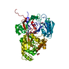 6lzqC 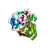 6lztC 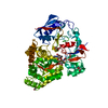 6lzuC 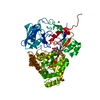 6lzvC  6lzwC  7ebmC  5yqwS C: citing same article ( S: Starting model for refinement |
|---|---|
| Similar structure data |


-Links
 PDBj
PDBj- Assembly
| Deposited unit | 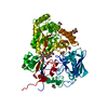
| ||||||||
|---|---|---|---|---|---|---|---|---|---|
| 1 |
| ||||||||
| Unit cell |
|
-Components
-Protein / Sugars , 2 types, 2 molecules A
| #1: Protein | Mass: 61288.059 Da / Num. of mol.: 1 Source method: isolated from a genetically manipulated source Source: (gene. exp.) Vibrio harveyi (strain 1DA3) (bacteria)Strain: 1DA3 / Gene: VME_26970 / Production host: |
|---|---|
| #2: Polysaccharide | 2-acetamido-2-deoxy-beta-D-glucopyranose-(1-4)-2-acetamido-2-deoxy-beta-D-glucopyranose |
-Non-polymers , 5 types, 329 molecules 


| #3: Chemical | ChemComp-EDO /  Mass: 62.068 Da / Num. of mol.: 13 / Source method: obtained synthetically / Formula: C2H6O2 Mass: 62.068 Da / Num. of mol.: 13 / Source method: obtained synthetically / Formula: C2H6O2#4: Chemical | ChemComp-MG /  Mass: 24.305 Da / Num. of mol.: 4 / Source method: obtained synthetically / Formula: Mg Mass: 24.305 Da / Num. of mol.: 4 / Source method: obtained synthetically / Formula: Mg#5: Chemical |  Mass: 40.078 Da / Num. of mol.: 2 / Source method: isolated from a natural source / Formula: Ca Mass: 40.078 Da / Num. of mol.: 2 / Source method: isolated from a natural source / Formula: Ca#6: Chemical |  Mass: 35.453 Da / Num. of mol.: 2 / Source method: obtained synthetically / Formula: Cl Mass: 35.453 Da / Num. of mol.: 2 / Source method: obtained synthetically / Formula: Cl#7: Water | ChemComp-HOH / | Mass: 18.015 Da / Num. of mol.: 308 / Source method: isolated from a natural source / Formula: H2O |
|---|
-Details
| Has ligand of interest | Y |
|---|---|
| Has protein modification | N |
-Experimental details
-Experiment
| Experiment | Method: X-RAY DIFFRACTION / Number of used crystals: 1 |
|---|
- Sample preparation
| Crystal | Density Matthews: 2.36 Å3/Da / Density % sol: 47.79 % |
|---|---|
| Crystal grow | Temperature: 293 K / Method: microbatch Details: 1.63mM chitotetramer, 0.06M Divalents (MgCl2, CaCl2), 0.1M Buffer System 1 (Imidazole, MES (acid)), pH 6.5, 30% v/v Precipitant Mix 2 (Ethylene glycol, PEG 8000) |
-Data collection
| Diffraction | Mean temperature: 100 K / Serial crystal experiment: N |
|---|---|
| Diffraction source | Source: SYNCHROTRON / Site: NSRRC  / Beamline: TPS 05A / Wavelength: 0.99984 Å / Beamline: TPS 05A / Wavelength: 0.99984 Å |
| Detector | Type: RAYONIX MX300-HS / Detector: CCD / Date: Dec 8, 2019 |
| Radiation | Protocol: SINGLE WAVELENGTH / Monochromatic (M) / Laue (L): M / Scattering type: x-ray |
| Radiation wavelength | Wavelength: 0.99984 Å / Relative weight: 1 |
| Reflection | Resolution: 1.5→50 Å / Num. obs: 89117 / % possible obs: 97.8 % / Redundancy: 3.4 % / Rpim(I) all: 0.04 / Rrim(I) all: 0.076 / Net I/σ(I): 19.99 |
| Reflection shell | Resolution: 1.5→1.53 Å / Redundancy: 2.3 % / Num. unique obs: 4293 / CC1/2: 0.762 / Rpim(I) all: 0.312 / Rrim(I) all: 0.506 / % possible all: 95.7 |
- Processing
| Software |
| |||||||||||||||||||||||||||||||||||||||||||||||||||||||||||||||||||||||||||||||||||||||||||||||||||||||||||||||||||||||||||||||||||||||||||||||||||||||||||||||||||||||||||||||||||||||||||||||||||||||||||||||||||||||||
|---|---|---|---|---|---|---|---|---|---|---|---|---|---|---|---|---|---|---|---|---|---|---|---|---|---|---|---|---|---|---|---|---|---|---|---|---|---|---|---|---|---|---|---|---|---|---|---|---|---|---|---|---|---|---|---|---|---|---|---|---|---|---|---|---|---|---|---|---|---|---|---|---|---|---|---|---|---|---|---|---|---|---|---|---|---|---|---|---|---|---|---|---|---|---|---|---|---|---|---|---|---|---|---|---|---|---|---|---|---|---|---|---|---|---|---|---|---|---|---|---|---|---|---|---|---|---|---|---|---|---|---|---|---|---|---|---|---|---|---|---|---|---|---|---|---|---|---|---|---|---|---|---|---|---|---|---|---|---|---|---|---|---|---|---|---|---|---|---|---|---|---|---|---|---|---|---|---|---|---|---|---|---|---|---|---|---|---|---|---|---|---|---|---|---|---|---|---|---|---|---|---|---|---|---|---|---|---|---|---|---|---|---|---|---|---|---|---|---|
| Refinement | Method to determine structure: MOLECULAR REPLACEMENT Starting model: 5yqw Resolution: 1.5→24.49 Å / SU ML: 0.13 / Cross valid method: FREE R-VALUE / σ(F): 1.36 / Phase error: 17.81 / Stereochemistry target values: ML
| |||||||||||||||||||||||||||||||||||||||||||||||||||||||||||||||||||||||||||||||||||||||||||||||||||||||||||||||||||||||||||||||||||||||||||||||||||||||||||||||||||||||||||||||||||||||||||||||||||||||||||||||||||||||||
| Solvent computation | Shrinkage radii: 0.9 Å / VDW probe radii: 1.11 Å / Solvent model: FLAT BULK SOLVENT MODEL | |||||||||||||||||||||||||||||||||||||||||||||||||||||||||||||||||||||||||||||||||||||||||||||||||||||||||||||||||||||||||||||||||||||||||||||||||||||||||||||||||||||||||||||||||||||||||||||||||||||||||||||||||||||||||
| Refinement step | Cycle: LAST / Resolution: 1.5→24.49 Å
| |||||||||||||||||||||||||||||||||||||||||||||||||||||||||||||||||||||||||||||||||||||||||||||||||||||||||||||||||||||||||||||||||||||||||||||||||||||||||||||||||||||||||||||||||||||||||||||||||||||||||||||||||||||||||
| Refine LS restraints |
| |||||||||||||||||||||||||||||||||||||||||||||||||||||||||||||||||||||||||||||||||||||||||||||||||||||||||||||||||||||||||||||||||||||||||||||||||||||||||||||||||||||||||||||||||||||||||||||||||||||||||||||||||||||||||
| LS refinement shell |
|
