- PDB-3e3k: Structural characterization of a putative endogenous metal chelat... -
+
Open data
ID or keywords:
Loading...
-
Basic information
Entry
Database: PDB / ID: 3e3k
Title
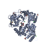
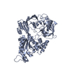
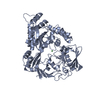
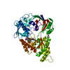
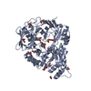
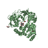
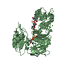
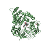
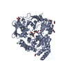
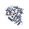
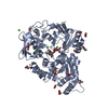
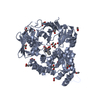
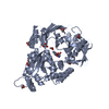
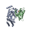
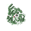
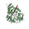
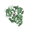
Structural characterization of a putative endogenous metal chelator in the periplasmic nickel transporter NikA (butane-1,2,4-tricarboxylate without nickel form)
Components
Nickel-binding periplasmic protein
Keywords
METAL TRANSPORT / Nickel / nickellophore / butane-1 / 2 / 4-tricarboxylate / transport
Function / homology
Function and homology information
nickel cation import across plasma membrane / metal cluster binding / nickel cation transport / peptide transport / peptide transmembrane transporter activity / negative chemotaxis / nickel cation binding / transition metal ion binding / ATP-binding cassette (ABC) transporter complex, substrate-binding subunit-containing / outer membrane-bounded periplasmic space ...nickel cation import across plasma membrane / metal cluster binding / nickel cation transport / peptide transport / peptide transmembrane transporter activity / negative chemotaxis / nickel cation binding / transition metal ion binding / ATP-binding cassette (ABC) transporter complex, substrate-binding subunit-containing / outer membrane-bounded periplasmic space / periplasmic space / heme binding / membrane Similarity search - Function
Nickel ABC transporter, substrate-binding protein NikA / Solute-binding protein family 5, conserved site / Bacterial extracellular solute-binding proteins, family 5 signature. / Dipeptide-binding Protein; domain 3 / Dipeptide-binding Protein; Domain 3 / Peptide/nickel binding protein, MppA-type / Solute-binding protein family 5 domain / Solute-binding protein family 5 / Bacterial extracellular solute-binding proteins, family 5 Middle / Periplasmic binding protein-like II ...Nickel ABC transporter, substrate-binding protein NikA / Solute-binding protein family 5, conserved site / Bacterial extracellular solute-binding proteins, family 5 signature. / Dipeptide-binding Protein; domain 3 / Dipeptide-binding Protein; Domain 3 / Peptide/nickel binding protein, MppA-type / Solute-binding protein family 5 domain / Solute-binding protein family 5 / Bacterial extracellular solute-binding proteins, family 5 Middle / Periplasmic binding protein-like II / D-Maltodextrin-Binding Protein; domain 2 / Roll / 3-Layer(aba) Sandwich / Alpha Beta Similarity search - Domain/homology
ACETATE ION / (2R)-butane-1,2,4-tricarboxylic acid / Nickel-binding periplasmic protein Similarity search - Component
In the structure databanks used in Yorodumi, some data are registered as the other names, "COVID-19 virus" and "2019-nCoV". Here are the details of the virus and the list of structure data.
Jan 31, 2019. EMDB accession codes are about to change! (news from PDBe EMDB page)
EMDB accession codes are about to change! (news from PDBe EMDB page)
The allocation of 4 digits for EMDB accession codes will soon come to an end. Whilst these codes will remain in use, new EMDB accession codes will include an additional digit and will expand incrementally as the available range of codes is exhausted. The current 4-digit format prefixed with “EMD-” (i.e. EMD-XXXX) will advance to a 5-digit format (i.e. EMD-XXXXX), and so on. It is currently estimated that the 4-digit codes will be depleted around Spring 2019, at which point the 5-digit format will come into force.
The EM Navigator/Yorodumi systems omit the EMD- prefix.
Related info.:Q: What is EMD? / ID/Accession-code notation in Yorodumi/EM Navigator
Yorodumi is a browser for structure data from EMDB, PDB, SASBDB, etc.
This page is also the successor to EM Navigator detail page, and also detail information page/front-end page for Omokage search.
The word "yorodu" (or yorozu) is an old Japanese word meaning "ten thousand". "mi" (miru) is to see.
Related info.:EMDB / PDB / SASBDB / Comparison of 3 databanks / Yorodumi Search / Aug 31, 2016. New EM Navigator & Yorodumi / Yorodumi Papers / Jmol/JSmol / Function and homology information / Changes in new EM Navigator and Yorodumi
 Movie
Movie Controller

 Yorodumi
 Open data
 Basic information
 Components
 Keywords
 Function and homology information
 X-RAY DIFFRACTION /
  Authors
 Citation
 Structure visualization
 Downloads & links
 Other downloads


 PDBj
PDBj Assembly
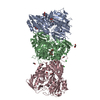


 Mass: 92.094 Da / Num. of mol.: 3 / Source method: obtained synthetically / Formula: C3H8O3
 Mass: 59.044 Da / Num. of mol.: 6 / Source method: obtained synthetically / Formula: C2H3O2
 Mass: 96.063 Da / Num. of mol.: 1 / Source method: obtained synthetically / Formula: SO4
 Mass: 190.151 Da / Num. of mol.: 3 / Source method: obtained synthetically / Formula: C7H10O6
 Mass: 35.453 Da / Num. of mol.: 4 / Source method: obtained synthetically / Formula: Cl
 Sample preparation
 / Beamline: ID14-3 / Wavelength: 0.931 Å
 Processing