 Movie
Movie Controller

[English] 日本語
 Yorodumi
- PDB-3mvy: X-ray structure of the diatomic oxo-intermediate NikA/1-Int', pri... -
+ Open data

- Basic information
| Entry | Database: PDB / ID: 3mvy | ||||||
|---|---|---|---|---|---|---|---|
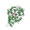
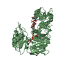
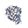
| Title | X-ray structure of the diatomic oxo-intermediate NikA/1-Int', prior hydroxylation | ||||||
 Components Components | Nickel-binding periplasmic protein | ||||||
 Keywords Keywords | TRANSPORT PROTEIN / Protein-bound iron complex | ||||||
| Function / homology |  Function and homology information Function and homology informationnickel cation import across plasma membrane / metal cluster binding / nickel cation transport / peptide transport / peptide transmembrane transporter activity / negative chemotaxis / nickel cation binding / transition metal ion binding / ATP-binding cassette (ABC) transporter complex / outer membrane-bounded periplasmic space ...nickel cation import across plasma membrane / metal cluster binding / nickel cation transport / peptide transport / peptide transmembrane transporter activity / negative chemotaxis / nickel cation binding / transition metal ion binding / ATP-binding cassette (ABC) transporter complex / outer membrane-bounded periplasmic space / periplasmic space / heme binding / membrane Similarity search - Function | ||||||
| Biological species |  | ||||||
| Method |  X-RAY DIFFRACTION / SYNCHROTRON / MOLECULAR REPLACEMENT / Resolution: 2.5 Å X-RAY DIFFRACTION / SYNCHROTRON / MOLECULAR REPLACEMENT / Resolution: 2.5 Å | ||||||
 Authors Authors | Cavazza, C. / Bochot, C. / Rousselot-Pailley, P. / Carpentier, P. / Cherrier, M.V. / Martin, L. / Marchi-Delapierre, C. / Fontecilla-Camps, J.C. / Menage, S. | ||||||
 Citation Citation | Journal: NAT.CHEM. / Year: 2010 Title: Crystallographic snapshots of the reaction of aromatic C-H with O(2) catalysed by a protein-bound iron complex Authors: Cavazza, C. / Bochot, C. / Rousselot-Pailley, P. / Carpentier, P. / Cherrier, M.V. / Martin, L. / Marchi-Delapierre, C. / Fontecilla-Camps, J.C. / Menage, S. | ||||||
| History |
|
- Structure visualization
| Structure viewer | Molecule: MolmilJmol/JSmol |
|---|
- Downloads & links
-Download
| PDBx/mmCIF format | 3mvy.cif.gz | 215.5 KB | Display | PDBx/mmCIF format |
|---|---|---|---|---|
| PDB format | pdb3mvy.ent.gz | 171.8 KB | Display | PDB format |
| PDBx/mmJSON format | 3mvy.json.gz | Tree view | PDBx/mmJSON format | |
| Others |  Other downloads Other downloads |
-Validation report
| Arichive directory | https://data.pdbj.org/pub/pdb/validation_reports/mv/3mvyftp://data.pdbj.org/pub/pdb/validation_reports/mv/3mvy | HTTPS FTP |
|---|
-Related structure data
| Related structure data |  3mvwC  3mvxC  3mvzC  3mw0C  3mz9C  3mzbC  1zlqS C: citing same article ( S: Starting model for refinement |
|---|---|
| Similar structure data |


-Links
 PDBj
PDBj- Assembly
| Deposited unit | 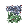
| ||||||||
|---|---|---|---|---|---|---|---|---|---|
| 1 | 
| ||||||||
| 2 | 
| ||||||||
| Unit cell |
|
-Components
-Protein , 1 types, 2 molecules AB
| #1: Protein | Mass: 56360.734 Da / Num. of mol.: 2 Source method: isolated from a genetically manipulated source Source: (gene. exp.) |
|---|
-Non-polymers , 8 types, 374 molecules 


| #2: Chemical | ChemComp-ACT /  Mass: 59.044 Da / Num. of mol.: 7 / Source method: obtained synthetically / Formula: C2H3O2 Mass: 59.044 Da / Num. of mol.: 7 / Source method: obtained synthetically / Formula: C2H3O2#3: Chemical | ChemComp-SO4 / |  Mass: 96.063 Da / Num. of mol.: 1 / Source method: obtained synthetically / Formula: SO4 Mass: 96.063 Da / Num. of mol.: 1 / Source method: obtained synthetically / Formula: SO4#4: Chemical | ChemComp-GOL /  Mass: 92.094 Da / Num. of mol.: 8 / Source method: obtained synthetically / Formula: C3H8O3 Mass: 92.094 Da / Num. of mol.: 8 / Source method: obtained synthetically / Formula: C3H8O3#5: Chemical |  Mass: 55.845 Da / Num. of mol.: 2 / Source method: obtained synthetically / Formula: Fe Mass: 55.845 Da / Num. of mol.: 2 / Source method: obtained synthetically / Formula: Fe#6: Chemical |  Mass: 372.415 Da / Num. of mol.: 2 / Source method: obtained synthetically / Formula: C20H24N2O5 Mass: 372.415 Da / Num. of mol.: 2 / Source method: obtained synthetically / Formula: C20H24N2O5#7: Chemical |  Mass: 31.999 Da / Num. of mol.: 2 / Source method: obtained synthetically / Formula: O2 Mass: 31.999 Da / Num. of mol.: 2 / Source method: obtained synthetically / Formula: O2#8: Chemical |  Mass: 35.453 Da / Num. of mol.: 2 / Source method: obtained synthetically / Formula: Cl Mass: 35.453 Da / Num. of mol.: 2 / Source method: obtained synthetically / Formula: Cl#9: Water | ChemComp-HOH / | Mass: 18.015 Da / Num. of mol.: 350 / Source method: isolated from a natural source / Formula: H2O |
|---|
-Experimental details
-Experiment
| Experiment | Method: X-RAY DIFFRACTION / Number of used crystals: 1 |
|---|
- Sample preparation
| Crystal | Density Matthews: 2.3 Å3/Da / Density % sol: 46.43 % |
|---|---|
| Crystal grow | Temperature: 293 K / Method: vapor diffusion, hanging drop / pH: 4.7 Details: 1.8M ammonium sulfate, 0.1M sodium acetate, pH 4.7, VAPOR DIFFUSION, HANGING DROP, temperature 293K |
-Data collection
| Diffraction source | Source: SYNCHROTRON / Site: ESRF  / Beamline: BM30A / Wavelength: 0.9796 Å / Beamline: BM30A / Wavelength: 0.9796 Å |
|---|---|
| Detector | Type: ADSC QUANTUM 315r / Detector: CCD / Date: Apr 15, 2009 / Details: Mirrors |
| Radiation | Protocol: SINGLE WAVELENGTH / Monochromatic (M) / Laue (L): M / Scattering type: x-ray |
| Radiation wavelength | Wavelength: 0.9796 Å / Relative weight: 1 |
| Reflection | Resolution: 2.5→43.5 Å / Num. obs: 34625 / % possible obs: 94 % / Redundancy: 2.3 % / Biso Wilson estimate: 36.9 Å2 / Rsym value: 0.106 / Net I/σ(I): 7.86 |
| Reflection shell | Highest resolution: 2.5 Å / Redundancy: 2.4 % / Mean I/σ(I) obs: 3.3 / Num. unique all: 6716 / Rsym value: 0.313 / % possible all: 88 |
- Processing
| Software | Name: REFMAC / Version: 5.5.0109 / Classification: refinement | ||||||||||||||||||||||||||||||||||||||||||||||||||||||||||||||||||||||||||||||||||||||||||||||||||||||||||||||||||||||||||||||||||||||||||||||||||||||||||||||||||||||||||
|---|---|---|---|---|---|---|---|---|---|---|---|---|---|---|---|---|---|---|---|---|---|---|---|---|---|---|---|---|---|---|---|---|---|---|---|---|---|---|---|---|---|---|---|---|---|---|---|---|---|---|---|---|---|---|---|---|---|---|---|---|---|---|---|---|---|---|---|---|---|---|---|---|---|---|---|---|---|---|---|---|---|---|---|---|---|---|---|---|---|---|---|---|---|---|---|---|---|---|---|---|---|---|---|---|---|---|---|---|---|---|---|---|---|---|---|---|---|---|---|---|---|---|---|---|---|---|---|---|---|---|---|---|---|---|---|---|---|---|---|---|---|---|---|---|---|---|---|---|---|---|---|---|---|---|---|---|---|---|---|---|---|---|---|---|---|---|---|---|---|---|---|
| Refinement | Method to determine structure: MOLECULAR REPLACEMENT Starting model: PDB ID 1ZLQ Resolution: 2.5→43.49 Å / Cor.coef. Fo:Fc: 0.942 / Cor.coef. Fo:Fc free: 0.882 / SU B: 9.515 / SU ML: 0.214 / Cross valid method: THROUGHOUT / ESU R: 1.176 / ESU R Free: 0.311 / Stereochemistry target values: MAXIMUM LIKELIHOOD / Details: HYDROGENS HAVE BEEN ADDED IN THE RIDING POSITIONS
| ||||||||||||||||||||||||||||||||||||||||||||||||||||||||||||||||||||||||||||||||||||||||||||||||||||||||||||||||||||||||||||||||||||||||||||||||||||||||||||||||||||||||||
| Solvent computation | Ion probe radii: 0.8 Å / Shrinkage radii: 0.8 Å / VDW probe radii: 1.4 Å / Solvent model: MASK | ||||||||||||||||||||||||||||||||||||||||||||||||||||||||||||||||||||||||||||||||||||||||||||||||||||||||||||||||||||||||||||||||||||||||||||||||||||||||||||||||||||||||||
| Displacement parameters | Biso mean: 21.989 Å2
| ||||||||||||||||||||||||||||||||||||||||||||||||||||||||||||||||||||||||||||||||||||||||||||||||||||||||||||||||||||||||||||||||||||||||||||||||||||||||||||||||||||||||||
| Refinement step | Cycle: LAST / Resolution: 2.5→43.49 Å
| ||||||||||||||||||||||||||||||||||||||||||||||||||||||||||||||||||||||||||||||||||||||||||||||||||||||||||||||||||||||||||||||||||||||||||||||||||||||||||||||||||||||||||
| Refine LS restraints |
| ||||||||||||||||||||||||||||||||||||||||||||||||||||||||||||||||||||||||||||||||||||||||||||||||||||||||||||||||||||||||||||||||||||||||||||||||||||||||||||||||||||||||||
| LS refinement shell | Resolution: 2.5→2.565 Å / Total num. of bins used: 20
|
