 Movie
Movie Controller

[English] 日本語
 Yorodumi
- PDB-7a0c: X-ray structure of NikA from Escherichia coli in complex with Fe-... -
+ Open data

- Basic information
| Entry | Database: PDB / ID: 7a0c | ||||||
|---|---|---|---|---|---|---|---|
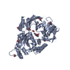
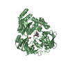
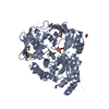
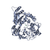
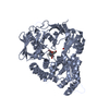
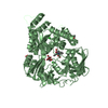
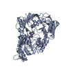
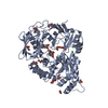
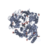
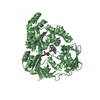
| Title | X-ray structure of NikA from Escherichia coli in complex with Fe-6-Me2-BPMCN | ||||||
 Components Components | Nickel-binding periplasmic protein | ||||||
 Keywords Keywords | METAL BINDING PROTEIN / Artificial metalloenzyme / cross-linked enzyme crystal / sulfoxidation | ||||||
| Function / homology |  Function and homology information Function and homology informationnickel cation import across plasma membrane / metal cluster binding / nickel cation transport / peptide transport / peptide transmembrane transporter activity / negative chemotaxis / nickel cation binding / transition metal ion binding / ATP-binding cassette (ABC) transporter complex, substrate-binding subunit-containing / outer membrane-bounded periplasmic space ...nickel cation import across plasma membrane / metal cluster binding / nickel cation transport / peptide transport / peptide transmembrane transporter activity / negative chemotaxis / nickel cation binding / transition metal ion binding / ATP-binding cassette (ABC) transporter complex, substrate-binding subunit-containing / outer membrane-bounded periplasmic space / periplasmic space / heme binding / membrane Similarity search - Function | ||||||
| Biological species |  | ||||||
| Method |  X-RAY DIFFRACTION / SYNCHROTRON / MOLECULAR REPLACEMENT / Resolution: 1.9 Å X-RAY DIFFRACTION / SYNCHROTRON / MOLECULAR REPLACEMENT / Resolution: 1.9 Å | ||||||
 Authors Authors | Cavazza, C. / Menage, S. | ||||||
| Funding support |  France, 1items France, 1items
| ||||||
 Citation Citation | Journal: Chemistry / Year: 2020 Title: A Selective Sulfide Oxidation Catalyzed by Heterogeneous Artificial Metalloenzymes Iron@NikA. Authors: Lopez, S. / Marchi-Delapierre, C. / Cavazza, C. / Menage, S. | ||||||
| History |
|
- Structure visualization
| Structure viewer | Molecule: MolmilJmol/JSmol |
|---|
- Downloads & links
-Download
| PDBx/mmCIF format | 7a0c.cif.gz | 231.6 KB | Display | PDBx/mmCIF format |
|---|---|---|---|---|
| PDB format | pdb7a0c.ent.gz | 183.6 KB | Display | PDB format |
| PDBx/mmJSON format | 7a0c.json.gz | Tree view | PDBx/mmJSON format | |
| Others |  Other downloads Other downloads |
-Validation report
| Arichive directory | https://data.pdbj.org/pub/pdb/validation_reports/a0/7a0cftp://data.pdbj.org/pub/pdb/validation_reports/a0/7a0c | HTTPS FTP |
|---|
-Related structure data
| Related structure data | 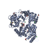 1zlqS S: Starting model for refinement |
|---|---|
| Similar structure data |
-Links
 PDBj
PDBj- Assembly
| Deposited unit | 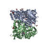
| ||||||||
|---|---|---|---|---|---|---|---|---|---|
| 1 | 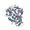
| ||||||||
| 2 | 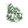
| ||||||||
| Unit cell |
|
-Components
-Protein , 1 types, 2 molecules AB
| #1: Protein | Mass: 56360.734 Da / Num. of mol.: 2 Source method: isolated from a genetically manipulated source Source: (gene. exp.) Strain: K12 / Gene: nikA, b3476, JW3441 / Production host: |
|---|
-Non-polymers , 7 types, 823 molecules 


| #2: Chemical |  Mass: 55.845 Da / Num. of mol.: 2 / Source method: obtained synthetically / Formula: Fe Mass: 55.845 Da / Num. of mol.: 2 / Source method: obtained synthetically / Formula: Fe#3: Chemical |  Mass: 396.526 Da / Num. of mol.: 2 / Source method: obtained synthetically / Formula: C23H32N4O2 Mass: 396.526 Da / Num. of mol.: 2 / Source method: obtained synthetically / Formula: C23H32N4O2#4: Chemical | ChemComp-ACT /  Mass: 59.044 Da / Num. of mol.: 9 / Source method: obtained synthetically / Formula: C2H3O2 Mass: 59.044 Da / Num. of mol.: 9 / Source method: obtained synthetically / Formula: C2H3O2#5: Chemical | ChemComp-GOL /  Mass: 92.094 Da / Num. of mol.: 9 / Source method: obtained synthetically / Formula: C3H8O3 Mass: 92.094 Da / Num. of mol.: 9 / Source method: obtained synthetically / Formula: C3H8O3#6: Chemical | ChemComp-MG /  Mass: 24.305 Da / Num. of mol.: 5 / Source method: obtained synthetically / Formula: Mg Mass: 24.305 Da / Num. of mol.: 5 / Source method: obtained synthetically / Formula: Mg#7: Chemical | ChemComp-CL / |  Mass: 35.453 Da / Num. of mol.: 1 / Source method: obtained synthetically / Formula: Cl Mass: 35.453 Da / Num. of mol.: 1 / Source method: obtained synthetically / Formula: Cl#8: Water | ChemComp-HOH / | Mass: 18.015 Da / Num. of mol.: 795 / Source method: isolated from a natural source / Formula: H2O |
|---|
-Details
| Has ligand of interest | N |
|---|
-Experimental details
-Experiment
| Experiment | Method: X-RAY DIFFRACTION / Number of used crystals: 1 |
|---|
- Sample preparation
| Crystal | Density Matthews: 2.24 Å3/Da / Density % sol: 45.17 % |
|---|---|
| Crystal grow | Temperature: 293 K / Method: vapor diffusion, hanging drop / pH: 4.6 Details: 1.7 M Ammonium sulfate, 100 mM sodium acetate pH 4.6 |
-Data collection
| Diffraction | Mean temperature: 93 K / Serial crystal experiment: N |
|---|---|
| Diffraction source | Source: SYNCHROTRON / Site: ESRF / Beamline: BM30A / Wavelength: 0.9799 Å |
| Detector | Type: ADSC QUANTUM 315r / Detector: CCD / Date: Jul 13, 2017 |
| Radiation | Protocol: SINGLE WAVELENGTH / Monochromatic (M) / Laue (L): M / Scattering type: x-ray |
| Radiation wavelength | Wavelength: 0.9799 Å / Relative weight: 1 |
| Reflection | Resolution: 1.9→43.875 Å / Num. obs: 80337 / % possible obs: 99.8 % / Redundancy: 5.3 % / CC1/2: 0.998 / Rsym value: 9.6 / Net I/σ(I): 13.06 |
| Reflection shell | Resolution: 1.9→2 Å / Num. unique obs: 11314 / CC1/2: 0.655 / Rsym value: 1.3 |
- Processing
| Software |
| ||||||||||||||||||||||||||||||||||||||||||||||||||||||||||||||||||||||||||||||||||||||||||||||||||||||||||||||||||||||||||||||||||||||||||||||||||||||||||||||||||||||||||||||||||||
|---|---|---|---|---|---|---|---|---|---|---|---|---|---|---|---|---|---|---|---|---|---|---|---|---|---|---|---|---|---|---|---|---|---|---|---|---|---|---|---|---|---|---|---|---|---|---|---|---|---|---|---|---|---|---|---|---|---|---|---|---|---|---|---|---|---|---|---|---|---|---|---|---|---|---|---|---|---|---|---|---|---|---|---|---|---|---|---|---|---|---|---|---|---|---|---|---|---|---|---|---|---|---|---|---|---|---|---|---|---|---|---|---|---|---|---|---|---|---|---|---|---|---|---|---|---|---|---|---|---|---|---|---|---|---|---|---|---|---|---|---|---|---|---|---|---|---|---|---|---|---|---|---|---|---|---|---|---|---|---|---|---|---|---|---|---|---|---|---|---|---|---|---|---|---|---|---|---|---|---|---|---|
| Refinement | Method to determine structure: MOLECULAR REPLACEMENT Starting model: 1ZLQ Resolution: 1.9→43.875 Å / SU ML: 0.24 / Cross valid method: THROUGHOUT / σ(F): 1.36 / Phase error: 23.28 / Stereochemistry target values: ML
| ||||||||||||||||||||||||||||||||||||||||||||||||||||||||||||||||||||||||||||||||||||||||||||||||||||||||||||||||||||||||||||||||||||||||||||||||||||||||||||||||||||||||||||||||||||
| Solvent computation | Shrinkage radii: 0.9 Å / VDW probe radii: 1.11 Å / Solvent model: FLAT BULK SOLVENT MODEL | ||||||||||||||||||||||||||||||||||||||||||||||||||||||||||||||||||||||||||||||||||||||||||||||||||||||||||||||||||||||||||||||||||||||||||||||||||||||||||||||||||||||||||||||||||||
| Displacement parameters | Biso max: 93.17 Å2 / Biso mean: 30.3509 Å2 / Biso min: 12.81 Å2 | ||||||||||||||||||||||||||||||||||||||||||||||||||||||||||||||||||||||||||||||||||||||||||||||||||||||||||||||||||||||||||||||||||||||||||||||||||||||||||||||||||||||||||||||||||||
| Refinement step | Cycle: final / Resolution: 1.9→43.875 Å
| ||||||||||||||||||||||||||||||||||||||||||||||||||||||||||||||||||||||||||||||||||||||||||||||||||||||||||||||||||||||||||||||||||||||||||||||||||||||||||||||||||||||||||||||||||||
| LS refinement shell | Refine-ID: X-RAY DIFFRACTION / Rfactor Rfree error: 0
|
