 Movie
Movie Controller

[English] 日本語
 Yorodumi
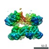
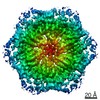
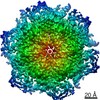
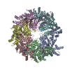
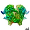
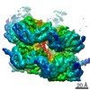
- PDB-7eav: The X-ray crystallographic structure of glycogen debranching enzy... -
+ Open data

- Basic information
| Entry | Database: PDB / ID: 7eav | ||||||||||||
|---|---|---|---|---|---|---|---|---|---|---|---|---|---|
| Title | The X-ray crystallographic structure of glycogen debranching enzyme from Sulfolobus solfataricus STB09 | ||||||||||||
 Components Components | Glycogen debranching enzyme | ||||||||||||
 Keywords Keywords | HYDROLASE / Glycogen Debranching Enzyme | ||||||||||||
| Function / homology |  Function and homology information Function and homology information: / glycogen catabolic process / aminopeptidase activity / hydrolase activity, hydrolyzing O-glycosyl compounds Similarity search - Function | ||||||||||||
| Biological species |   Saccharolobus solfataricus (archaea) Saccharolobus solfataricus (archaea) | ||||||||||||
| Method |  X-RAY DIFFRACTION / SYNCHROTRON / MOLECULAR REPLACEMENT / Resolution: 2.803 Å X-RAY DIFFRACTION / SYNCHROTRON / MOLECULAR REPLACEMENT / Resolution: 2.803 Å | ||||||||||||
 Authors Authors | Li, Z.F. / Ban, X.F. / Tian, Y.X. / Li, C.M. / Cheng, L. / Hong, Y. / Gu, Z.B. | ||||||||||||
| Funding support |  China, 3items China, 3items
| ||||||||||||
 Citation Citation | Journal: To Be Published Title: The X-ray Crystallographic Structure of Debranching Enzyme from Sulfolobus solfataricus STB09 Authors: Li, Z.F. / Ban, X.F. / Tian, Y.X. / Li, C.M. / Gu, Z.B. | ||||||||||||
| History |
|
- Structure visualization
| Structure viewer | Molecule: MolmilJmol/JSmol |
|---|
- Downloads & links
-Download
| PDBx/mmCIF format | 7eav.cif.gz | 296.4 KB | Display | PDBx/mmCIF format |
|---|---|---|---|---|
| PDB format | pdb7eav.ent.gz | 240.3 KB | Display | PDB format |
| PDBx/mmJSON format | 7eav.json.gz | Tree view | PDBx/mmJSON format | |
| Others |  Other downloads Other downloads |
-Validation report
| Summary document | 7eav_validation.pdf.gz | 442.4 KB | Display | wwPDB validaton report |
|---|---|---|---|---|
| Full document | 7eav_full_validation.pdf.gz | 478.3 KB | Display | |
| Data in XML | 7eav_validation.xml.gz | 54.3 KB | Display | |
| Data in CIF | 7eav_validation.cif.gz | 73.4 KB | Display | |
| Arichive directory | https://data.pdbj.org/pub/pdb/validation_reports/ea/7eavftp://data.pdbj.org/pub/pdb/validation_reports/ea/7eav | HTTPS FTP |
-Related structure data
| Related structure data |  2vr5S S: Starting model for refinement |
|---|---|
| Similar structure data |

-Links
 PDBj
PDBj

- Assembly
| Deposited unit | 
| ||||||||
|---|---|---|---|---|---|---|---|---|---|
| 1 | 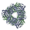
| ||||||||
| Unit cell |
|
-Components
| #1: Protein | Mass: 83181.859 Da / Num. of mol.: 2 Source method: isolated from a genetically manipulated source Source: (gene. exp.) Saccharolobus solfataricus (archaea)Gene: orf c06021, glgX, SSOP1_2225, SULA_2828, SULB_2829, SULC_2826, SULG_14405, SULH_14405, SULI_14405, SULM_14335, SULN_14325, SULO_14345, SULZ_14425 Production host:  #2: Water | ChemComp-HOH / |  Mass: 18.015 Da / Num. of mol.: 180 / Source method: isolated from a natural source / Formula: H2O Mass: 18.015 Da / Num. of mol.: 180 / Source method: isolated from a natural source / Formula: H2OHas protein modification | Y | |
|---|
-Experimental details
-Experiment
| Experiment | Method: X-RAY DIFFRACTION / Number of used crystals: 1 |
|---|
- Sample preparation
| Crystal | Density Matthews: 3.23 Å3/Da / Density % sol: 61.88 % |
|---|---|
| Crystal grow | Temperature: 291 K / Method: vapor diffusion, hanging drop / Details: PEG3000,lithium sulfate, imidazole |
-Data collection
| Diffraction | Mean temperature: 100 K / Serial crystal experiment: N |
|---|---|
| Diffraction source | Source: SYNCHROTRON / Site: SSRF / Beamline: BL19U1 / Wavelength: 0.9785 Å |
| Detector | Type: DECTRIS PILATUS3 6M / Detector: PIXEL / Date: Jun 25, 2020 |
| Radiation | Protocol: SINGLE WAVELENGTH / Monochromatic (M) / Laue (L): M / Scattering type: x-ray |
| Radiation wavelength | Wavelength: 0.9785 Å / Relative weight: 1 |
| Reflection | Resolution: 2.8→50 Å / Num. obs: 52466 / % possible obs: 99.9 % / Redundancy: 11.8 % / Rpim(I) all: 0.162 / Net I/σ(I): 6.4 |
| Reflection shell | Resolution: 2.8→2.89 Å / Num. unique obs: 4530 / CC1/2: 0.595 |
- Processing
| Software |
| ||||||||||||||||||||||||||||||||||||||||||||||||||||||||||||||||||||||||||||||||||||||||||||||||||||||||||||||||||
|---|---|---|---|---|---|---|---|---|---|---|---|---|---|---|---|---|---|---|---|---|---|---|---|---|---|---|---|---|---|---|---|---|---|---|---|---|---|---|---|---|---|---|---|---|---|---|---|---|---|---|---|---|---|---|---|---|---|---|---|---|---|---|---|---|---|---|---|---|---|---|---|---|---|---|---|---|---|---|---|---|---|---|---|---|---|---|---|---|---|---|---|---|---|---|---|---|---|---|---|---|---|---|---|---|---|---|---|---|---|---|---|---|---|---|---|
| Refinement | Method to determine structure: MOLECULAR REPLACEMENT Starting model: 2vr5 Resolution: 2.803→49.113 Å / SU ML: 0.73 / Cross valid method: THROUGHOUT / σ(F): 1.35 / Phase error: 42.25 / Stereochemistry target values: ML
| ||||||||||||||||||||||||||||||||||||||||||||||||||||||||||||||||||||||||||||||||||||||||||||||||||||||||||||||||||
| Solvent computation | Shrinkage radii: 0.9 Å / VDW probe radii: 1.11 Å / Solvent model: FLAT BULK SOLVENT MODEL | ||||||||||||||||||||||||||||||||||||||||||||||||||||||||||||||||||||||||||||||||||||||||||||||||||||||||||||||||||
| Displacement parameters | Biso max: 118.36 Å2 / Biso mean: 61.0181 Å2 / Biso min: 23.33 Å2 | ||||||||||||||||||||||||||||||||||||||||||||||||||||||||||||||||||||||||||||||||||||||||||||||||||||||||||||||||||
| Refinement step | Cycle: final / Resolution: 2.803→49.113 Å
| ||||||||||||||||||||||||||||||||||||||||||||||||||||||||||||||||||||||||||||||||||||||||||||||||||||||||||||||||||
| LS refinement shell | Refine-ID: X-RAY DIFFRACTION / Rfactor Rfree error: 0
|
