 Movie
Movie Controller

[English] 日本語
 Yorodumi
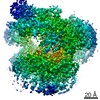
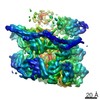
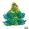
- PDB-2vuy: Crystal structure of Glycogen Debranching exzyme TreX from Sulfol... -
+ Open data

- Basic information
| Entry | Database: PDB / ID: 2vuy | ||||||
|---|---|---|---|---|---|---|---|
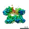
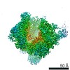
| Title | Crystal structure of Glycogen Debranching exzyme TreX from Sulfolobus solfatarius | ||||||
 Components Components | GLYCOGEN OPERON PROTEIN GLGX | ||||||
 Keywords Keywords | HYDROLASE / GLYCOSIDASE / GLYCOSYL HYDROLASE | ||||||
| Function / homology |  Function and homology information Function and homology informationamylo-alpha-1,6-glucosidase activity / glycogen catabolic process / aminopeptidase activity Similarity search - Function | ||||||
| Biological species |   SULFOLOBUS SOLFATARICUS (archaea) SULFOLOBUS SOLFATARICUS (archaea) | ||||||
| Method |  X-RAY DIFFRACTION / SYNCHROTRON / MOLECULAR REPLACEMENT / Resolution: 3 Å X-RAY DIFFRACTION / SYNCHROTRON / MOLECULAR REPLACEMENT / Resolution: 3 Å | ||||||
 Authors Authors | Song, H.-N. / Yoon, S.-M. / Cha, H.-J. / Park, K.-H. / Woo, E.-J. | ||||||
 Citation Citation | Journal: J.Biol.Chem. / Year: 2008 Title: Structural Insight Into the Bifunctional Mechanism of the Glycogen-Debranching Enzyme Trex from the Archaeon Sulfolobus Solfataricus. Authors: Woo, E. / Lee, S. / Cha, H. / Park, J. / Yoon, S. / Song, H. / Park, K. | ||||||
| History |
| ||||||
| Remark 700 | SHEET DETERMINATION METHOD: DSSP THE SHEETS PRESENTED AS "AD", "BD" IN EACH CHAIN ON SHEET RECORDS ... SHEET DETERMINATION METHOD: DSSP THE SHEETS PRESENTED AS "AD", "BD" IN EACH CHAIN ON SHEET RECORDS BELOW IS ACTUALLY AN 8-STRANDED BARREL THIS IS REPRESENTED BY A 9-STRANDED SHEET IN WHICH THE FIRST AND LAST STRANDS ARE IDENTICAL. |
- Structure visualization
| Structure viewer | Molecule: MolmilJmol/JSmol |
|---|
- Downloads & links
-Download
| PDBx/mmCIF format | 2vuy.cif.gz | 287.3 KB | Display | PDBx/mmCIF format |
|---|---|---|---|---|
| PDB format | pdb2vuy.ent.gz | 235.1 KB | Display | PDB format |
| PDBx/mmJSON format | 2vuy.json.gz | Tree view | PDBx/mmJSON format | |
| Others |  Other downloads Other downloads |
-Validation report
| Arichive directory | https://data.pdbj.org/pub/pdb/validation_reports/vu/2vuyftp://data.pdbj.org/pub/pdb/validation_reports/vu/2vuy | HTTPS FTP |
|---|
-Related structure data
| Related structure data |  2vncC  2vr5C  1bf2S C: citing same article ( S: Starting model for refinement |
|---|---|
| Similar structure data |


-Links
 PDBj
PDBj

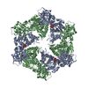
- Assembly
| Deposited unit | 
| ||||||||
|---|---|---|---|---|---|---|---|---|---|
| 1 | 
| ||||||||
| Unit cell |
|
-Components
| #1: Protein | Mass: 83181.859 Da / Num. of mol.: 2 Source method: isolated from a genetically manipulated source Details: DISULFIDE BOND BETWEEN A254 AND A261, A505 AND A519, B254 AND B261, B505 AND B519 Source: (gene. exp.) SULFOLOBUS SOLFATARICUS (archaea) / Production host:  References: UniProt: P95868, Hydrolases; Glycosylases; Glycosidases, i.e. enzymes that hydrolyse O- and S-glycosyl compounds Has protein modification | Y | |
|---|
-Experimental details
-Experiment
| Experiment | Method: X-RAY DIFFRACTION / Number of used crystals: 1 |
|---|
- Sample preparation
| Crystal | Density Matthews: 3.19 Å3/Da / Density % sol: 61.4 % / Description: NONE |
|---|---|
| Crystal grow | pH: 9.5 / Details: 16% PEG 8000, 0.2M NACL, 0.1 CHESS BUFFER(PH9.5) |
-Data collection
| Diffraction | Mean temperature: 95 K |
|---|---|
| Diffraction source | Source: SYNCHROTRON / Site: PAL/PLS  / Beamline: 4A / Wavelength: 1 / Beamline: 4A / Wavelength: 1 |
| Detector | Type: ADSC CCD / Detector: CCD / Date: Feb 20, 2006 |
| Radiation | Protocol: SINGLE WAVELENGTH / Monochromatic (M) / Laue (L): M / Scattering type: x-ray |
| Radiation wavelength | Wavelength: 1 Å / Relative weight: 1 |
| Reflection | Resolution: 3→50 Å / Num. obs: 39857 / % possible obs: 98.4 % / Observed criterion σ(I): 0 / Redundancy: 10.5 % / Rmerge(I) obs: 0.08 / Net I/σ(I): 12.8 |
| Reflection shell | Resolution: 3→3.11 Å / Redundancy: 9.2 % / Rmerge(I) obs: 0.24 / % possible all: 95.8 |
- Processing
| Software | Name: PHASER / Classification: phasing | ||||||||||||||||||||||||||||||||||||||||||||||||||||||||||||
|---|---|---|---|---|---|---|---|---|---|---|---|---|---|---|---|---|---|---|---|---|---|---|---|---|---|---|---|---|---|---|---|---|---|---|---|---|---|---|---|---|---|---|---|---|---|---|---|---|---|---|---|---|---|---|---|---|---|---|---|---|---|
| Refinement | Method to determine structure: MOLECULAR REPLACEMENT Starting model: PDB ENTRY 1BF2 Resolution: 3→29.86 Å / Rfactor Rfree error: 0.005 / Isotropic thermal model: RESTRAINED / Cross valid method: THROUGHOUT / σ(F): 0 / Details: BULK SOLVENT MODEL USED
| ||||||||||||||||||||||||||||||||||||||||||||||||||||||||||||
| Displacement parameters | Biso mean: 35.5 Å2
| ||||||||||||||||||||||||||||||||||||||||||||||||||||||||||||
| Refine analyze |
| ||||||||||||||||||||||||||||||||||||||||||||||||||||||||||||
| Refinement step | Cycle: LAST / Resolution: 3→29.86 Å
| ||||||||||||||||||||||||||||||||||||||||||||||||||||||||||||
| Refine LS restraints |
| ||||||||||||||||||||||||||||||||||||||||||||||||||||||||||||
| LS refinement shell | Resolution: 3→3.19 Å / Rfactor Rfree error: 0.019 / Total num. of bins used: 6
| ||||||||||||||||||||||||||||||||||||||||||||||||||||||||||||
| Xplor file | Serial no: 1 / Param file: PROTEIN_REP.PARAM / Topol file: PROTEIN.TOP |
