[English] 日本語
Yorodumi
- EMDB-23776: Structure of human p97 ATPase L464P mutant -

+
Open data


ID or keywords:

Loading...

-
Basic information

Entry
Database: EMDB / ID: EMD-23776
TitleStructure of human p97 ATPase L464P mutant
Map dataState B (symmetrized)
Sample
  • Complex: Human p97 L464P mutant
    • Protein or peptide: Transitional endoplasmic reticulum ATPase
  • Ligand: ADENOSINE-5'-DIPHOSPHATE
Function / homology
Function and homology information


positive regulation of Lys63-specific deubiquitinase activity / flavin adenine dinucleotide catabolic process / positive regulation of oxidative phosphorylation / VCP-NSFL1C complex / protein-DNA covalent cross-linking repair / endosome to lysosome transport via multivesicular body sorting pathway / endoplasmic reticulum stress-induced pre-emptive quality control / cellular response to arsenite ion / BAT3 complex binding / Derlin-1 retrotranslocation complex ...positive regulation of Lys63-specific deubiquitinase activity / flavin adenine dinucleotide catabolic process / positive regulation of oxidative phosphorylation / VCP-NSFL1C complex / protein-DNA covalent cross-linking repair / endosome to lysosome transport via multivesicular body sorting pathway / endoplasmic reticulum stress-induced pre-emptive quality control / cellular response to arsenite ion / BAT3 complex binding / Derlin-1 retrotranslocation complex / positive regulation of protein K63-linked deubiquitination / deubiquitinase activator activity / mitotic spindle disassembly / VCP-NPL4-UFD1 AAA ATPase complex / aggresome assembly / regulation of protein localization to chromatin / NADH metabolic process / vesicle-fusing ATPase / cellular response to misfolded protein / : / positive regulation of mitochondrial membrane potential / stress granule disassembly / K48-linked polyubiquitin modification-dependent protein binding / negative regulation of protein localization to chromatin / ERAD pathway / ubiquitin-modified protein reader activity / retrograde protein transport, ER to cytosol / regulation of aerobic respiration / ATPase complex / regulation of synapse organization / positive regulation of ATP biosynthetic process / ubiquitin-specific protease binding / ubiquitin-like protein ligase binding / autophagosome maturation / RHOH GTPase cycle / polyubiquitin modification-dependent protein binding / HSF1 activation / translesion synthesis / endoplasmic reticulum to Golgi vesicle-mediated transport / MHC class I protein binding / Protein methylation / interstrand cross-link repair / negative regulation of smoothened signaling pathway / Attachment and Entry / ATP metabolic process / : / endoplasmic reticulum unfolded protein response / proteasome complex / lipid droplet / viral genome replication / Josephin domain DUBs / N-glycan trimming in the ER and Calnexin/Calreticulin cycle / ADP binding / proteasomal protein catabolic process / Hh mutants are degraded by ERAD / positive regulation of protein-containing complex assembly / Defective CFTR causes cystic fibrosis / Hedgehog ligand biogenesis / macroautophagy / Translesion Synthesis by POLH / ABC-family proteins mediated transport / establishment of protein localization / autophagy / Aggrephagy / cytoplasmic stress granule / positive regulation of non-canonical NF-kappaB signal transduction / positive regulation of canonical Wnt signaling pathway / positive regulation of protein catabolic process / activation of cysteine-type endopeptidase activity involved in apoptotic process / double-strand break repair / KEAP1-NFE2L2 pathway / azurophil granule lumen / Ovarian tumor domain proteases / positive regulation of proteasomal ubiquitin-dependent protein catabolic process / E3 ubiquitin ligases ubiquitinate target proteins / site of double-strand break / cellular response to heat / Neddylation / ubiquitin-dependent protein catabolic process / proteasome-mediated ubiquitin-dependent protein catabolic process / protein phosphatase binding / regulation of apoptotic process / secretory granule lumen / ficolin-1-rich granule lumen / Attachment and Entry / protein ubiquitination / protein domain specific binding / DNA repair / intracellular membrane-bounded organelle / lipid binding / glutamatergic synapse / DNA damage response / ubiquitin protein ligase binding / Neutrophil degranulation / endoplasmic reticulum membrane / perinuclear region of cytoplasm / endoplasmic reticulum / ATP hydrolysis activity / protein-containing complex / RNA binding
Similarity search - Function
AAA ATPase, CDC48 family / Cell division protein 48 (CDC48), N-terminal domain / CDC48, N-terminal subdomain / Cell division protein 48 (CDC48) N-terminal domain / CDC48, domain 2 / Cell division protein 48 (CDC48), domain 2 / Cell division protein 48 (CDC48) domain 2 / CDC48 domain 2-like superfamily / Aspartate decarboxylase-like domain superfamily / AAA ATPase, AAA+ lid domain ...AAA ATPase, CDC48 family / Cell division protein 48 (CDC48), N-terminal domain / CDC48, N-terminal subdomain / Cell division protein 48 (CDC48) N-terminal domain / CDC48, domain 2 / Cell division protein 48 (CDC48), domain 2 / Cell division protein 48 (CDC48) domain 2 / CDC48 domain 2-like superfamily / Aspartate decarboxylase-like domain superfamily / AAA ATPase, AAA+ lid domain / AAA+ lid domain / ATPase, AAA-type, conserved site / AAA-protein family signature. / ATPase family associated with various cellular activities (AAA) / ATPase, AAA-type, core / ATPases associated with a variety of cellular activities / AAA+ ATPase domain / P-loop containing nucleoside triphosphate hydrolase
Similarity search - Domain/homology
Transitional endoplasmic reticulum ATPase
Similarity search - Component
Biological speciesHomo sapiens (human)
Methodsingle particle reconstruction / cryo EM / Resolution: 4.12 Å
AuthorsZhang X / Gui L / Li S / Nandi P / Columbres RC / Wong DE / Moen DR / Lin HJ / Chiu P-L / Chou T-F
Funding support United States, 2 items
OrganizationGrant numberCountry
National Institutes of Health/National Institute of Neurological Disorders and Stroke (NIH/NINDS)R01NS100815 United States
National Institutes of Health/National Institute of Neurological Disorders and Stroke (NIH/NINDS)R01NS102279 United States
Citation
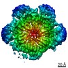
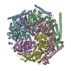
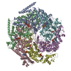
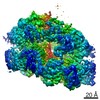
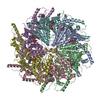
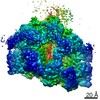
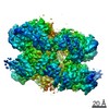
Journal: Biochem J / Year: 2021
Title: Conserved L464 in p97 D1-D2 linker is critical for p97 cofactor regulated ATPase activity.
Authors: Xiaoyi Zhang / Lin Gui / Shan Li / Purbasha Nandi / Rod Carlo Columbres / Daniel E Wong / Derek R Moen / Henry J Lin / Po-Lin Chiu / Tsui-Fen Chou /
Abstract: p97 protein is a highly conserved, abundant, functionally diverse, structurally dynamic homohexameric AAA enzyme-containing N, D1, and D2 domains. A truncated p97 protein containing the N and D1 ...p97 protein is a highly conserved, abundant, functionally diverse, structurally dynamic homohexameric AAA enzyme-containing N, D1, and D2 domains. A truncated p97 protein containing the N and D1 domains and the D1-D2 linker (ND1L) exhibits 79% of wild-type (WT) ATPase activity whereas the ND1 domain alone without the linker only has 2% of WT activity. To investigate the relationship between the D1-D2 linker and the D1 domain, we produced p97 ND1L mutants and demonstrated that this 22-residue linker region is essential for D1 ATPase activity. The conserved amino acid leucine 464 (L464) is critical for regulating D1 and D2 ATPase activity by p97 cofactors p37, p47, and Npl4-Ufd1 (NU). Changing leucine to alanine, proline, or glutamate increased the maximum rate of ATP turnover (kcat) of p47-regulated ATPase activities for these mutants, but not for WT. p37 and p47 increased the kcat of the proline substituted linker, suggesting that they induced linker conformations facilitating ATP hydrolysis. NU inhibited D1 ATPase activities of WT and mutant ND1L proteins, but activated D2 ATPase activity of full-length p97. To further understand the mutant mechanism, we used single-particle cryo-EM to visualize the full-length p97L464P and revealed the conformational change of the D1-D2 linker, resulting in a movement of the helix-turn-helix motif (543-569). Taken together with the biochemical and structural results we conclude that the linker helps maintain D1 in a competent conformation and relays the communication to/from the N-domain to the D1 and D2 ATPase domains, which are ∼50 Å away.
History
DepositionApr 5, 2021-
Header (metadata) releaseAug 25, 2021-
Map releaseAug 25, 2021-
UpdateSep 22, 2021-
Current statusSep 22, 2021Processing site: RCSB / Status: Released

-
Structure visualization

Movie
  • Surface view with section colored by density value
  • Surface level: 0.402
  • Imaged by UCSF Chimera
  • Download
  • Surface view colored by cylindrical radius
  • Surface level: 0.402
  • Imaged by UCSF Chimera
  • Download
  • Surface view with fitted model
  • Atomic models: PDB-7mdo
  • Surface level: 0.402
  • Imaged by UCSF Chimera
  • Download
Movie viewer
Structure viewerEM map:
SurfViewMolmilJmol/JSmol
Supplemental images

Downloads & links

-
Map

FileDownload / File: emd_23776.map.gz / Format: CCP4 / Size: 52.7 MB / Type: IMAGE STORED AS FLOATING POINT NUMBER (4 BYTES)
AnnotationState B (symmetrized)
Voxel sizeX=Y=Z: 1.56 Å
Density
Contour LevelBy AUTHOR: 0.402 / Movie #1: 0.402
Minimum - Maximum-1.279128 - 2.887218
Average (Standard dev.)-0.0011680258 (±0.07250735)
SymmetrySpace group: 1
Details

EMDB XML:

Map geometry
Axis orderXYZ
Origin000
Dimensions240240240
Spacing240240240
CellA=B=C: 374.4 Å
α=β=γ: 90.0 °

CCP4 map header:

modeImage stored as Reals
Å/pix. X/Y/Z1.561.561.56
M x/y/z240240240
origin x/y/z0.0000.0000.000
length x/y/z374.400374.400374.400
α/β/γ90.00090.00090.000
MAP C/R/S123
start NC/NR/NS000
NC/NR/NS240240240
D min/max/mean-1.2792.887-0.001

-
Supplemental data

-
Sample components

-
Entire : Human p97 L464P mutant

EntireName: Human p97 L464P mutant
Components
  • Complex: Human p97 L464P mutant
    • Protein or peptide: Transitional endoplasmic reticulum ATPase
  • Ligand: ADENOSINE-5'-DIPHOSPHATE

-
Supramolecule #1: Human p97 L464P mutant

SupramoleculeName: Human p97 L464P mutant / type: complex / ID: 1 / Parent: 0 / Macromolecule list: #1
Source (natural)Organism: Homo sapiens (human)
Recombinant expressionOrganism: Escherichia coli (E. coli)
Molecular weightTheoretical: 536 KDa

-
Macromolecule #1: Transitional endoplasmic reticulum ATPase

MacromoleculeName: Transitional endoplasmic reticulum ATPase / type: protein_or_peptide / ID: 1 / Number of copies: 6 / Enantiomer: LEVO / EC number: vesicle-fusing ATPase
Source (natural)Organism: Homo sapiens (human)
Molecular weightTheoretical: 89.420773 KDa
Recombinant expressionOrganism: Escherichia coli (E. coli)
SequenceString: MASGADSKGD DLSTAILKQK NRPNRLIVDE AINEDNSVVS LSQPKMDELQ LFRGDTVLLK GKKRREAVCI VLSDDTCSDE KIRMNRVVR NNLRVRLGDV ISIQPCPDVK YGKRIHVLPI DDTVEGITGN LFEVYLKPYF LEAYRPIRKG DIFLVRGGMR A VEFKVVET ...String:
MASGADSKGD DLSTAILKQK NRPNRLIVDE AINEDNSVVS LSQPKMDELQ LFRGDTVLLK GKKRREAVCI VLSDDTCSDE KIRMNRVVR NNLRVRLGDV ISIQPCPDVK YGKRIHVLPI DDTVEGITGN LFEVYLKPYF LEAYRPIRKG DIFLVRGGMR A VEFKVVET DPSPYCIVAP DTVIHCEGEP IKREDEEESL NEVGYDDIGG CRKQLAQIKE MVELPLRHPA LFKAIGVKPP RG ILLYGPP GTGKTLIARA VANETGAFFF LINGPEIMSK LAGESESNLR KAFEEAEKNA PAIIFIDELD AIAPKREKTH GEV ERRIVS QLLTLMDGLK QRAHVIVMAA TNRPNSIDPA LRRFGRFDRE VDIGIPDATG RLEILQIHTK NMKLADDVDL EQVA NETHG HVGADLAALC SEAALQAIRK KMDLIDLEDE TIDAEVMNSL AVTMDDFRWA LSQSNPSAPR ETVVEVPQVT WEDIG GLED VKRELQELVQ YPVEHPDKFL KFGMTPSKGV LFYGPPGCGK TLLAKAIANE CQANFISIKG PELLTMWFGE SEANVR EIF DKARQAAPCV LFFDELDSIA KARGGNIGDG GGAADRVINQ ILTEMDGMST KKNVFIIGAT NRPDIIDPAI LRPGRLD QL IYIPLPDEKS RVAILKANLR KSPVAKDVDL EFLAKMTNGF SGADLTEICQ RACKLAIRES IESEIRRERE RQTNPSAM E VEEDDPVPEI RRDHFEEAMR FARRSVSDND IRKYEMFAQT LQQSRGFGSF RFPSGNQGGA GPSQGSGGGT GGSVYTEDN DDDLYG

-
Macromolecule #2: ADENOSINE-5'-DIPHOSPHATE

MacromoleculeName: ADENOSINE-5'-DIPHOSPHATE / type: ligand / ID: 2 / Number of copies: 6 / Formula: ADP
Molecular weightTheoretical: 427.201 Da
Chemical component information

ChemComp-ADP:
ADENOSINE-5'-DIPHOSPHATE / ADP, energy-carrying molecule*YM / Adenosine diphosphate

-
Experimental details

-
Structure determination

Methodcryo EM
Processingsingle particle reconstruction
Aggregation stateparticle

-
Sample preparation

BufferpH: 7.4
GridModel: C-flat-2/1 / Material: COPPER / Support film - Material: CARBON / Support film - topology: HOLEY ARRAY / Pretreatment - Type: GLOW DISCHARGE
VitrificationCryogen name: ETHANE

-
Electron microscopy

MicroscopeFEI TITAN KRIOS
Electron beamAcceleration voltage: 300 kV / Electron source: FIELD EMISSION GUN
Electron opticsC2 aperture diameter: 70.0 µm / Illumination mode: FLOOD BEAM / Imaging mode: BRIGHT FIELDBright-field microscopy / Cs: 2.7 mm
Sample stageSpecimen holder model: FEI TITAN KRIOS AUTOGRID HOLDER / Cooling holder cryogen: NITROGEN
Image recordingFilm or detector model: GATAN K2 SUMMIT (4k x 4k) / Detector mode: COUNTING / Average electron dose: 64.8 e/Å2
Experimental equipment
Model: Titan Krios / Image courtesy: FEI Company

-
Image processing

CTF correctionSoftware - Name: cryoSPARC (ver. 3.1)
Initial angle assignmentType: MAXIMUM LIKELIHOOD / Software - Name: cryoSPARC (ver. 3.1)
Final angle assignmentType: MAXIMUM LIKELIHOOD / Software - Name: cryoSPARC (ver. 3.1)
Final reconstructionApplied symmetry - Point group: C6 (6 fold cyclic) / Algorithm: BACK PROJECTION / Resolution.type: BY AUTHOR / Resolution: 4.12 Å / Resolution method: FSC 0.143 CUT-OFF / Software - Name: cryoSPARC (ver. 3.1) / Number images used: 81865

-
Atomic model buiding 1

Initial modelPDB ID:
RefinementProtocol: RIGID BODY FIT
Output model

PDB-7mdo:
Structure of human p97 ATPase L464P mutant

+
About Yorodumi

-
News

-
Feb 9, 2022. New format data for meta-information of EMDB entries

New format data for meta-information of EMDB entries

  • Version 3 of the EMDB header file is now the official format.
  • The previous official version 1.9 will be removed from the archive.

Related info.:EMDB header

External links:wwPDB to switch to version 3 of the EMDB data model

-
Aug 12, 2020. Covid-19 info

Covid-19 info

URL: https://pdbj.org/emnavi/covid19.php

New page: Covid-19 featured information page in EM Navigator.

Related info.:Covid-19 info / Mar 5, 2020. Novel coronavirus structure data

+
Mar 5, 2020. Novel coronavirus structure data

Novel coronavirus structure data

Related info.:Yorodumi Speices / Aug 12, 2020. Covid-19 info

External links:COVID-19 featured content - PDBj / Molecule of the Month (242):Coronavirus Proteases

+
Jan 31, 2019. EMDB accession codes are about to change! (news from PDBe EMDB page)

EMDB accession codes are about to change! (news from PDBe EMDB page)

  • The allocation of 4 digits for EMDB accession codes will soon come to an end. Whilst these codes will remain in use, new EMDB accession codes will include an additional digit and will expand incrementally as the available range of codes is exhausted. The current 4-digit format prefixed with “EMD-” (i.e. EMD-XXXX) will advance to a 5-digit format (i.e. EMD-XXXXX), and so on. It is currently estimated that the 4-digit codes will be depleted around Spring 2019, at which point the 5-digit format will come into force.
  • The EM Navigator/Yorodumi systems omit the EMD- prefix.

Related info.:Q: What is EMD? / ID/Accession-code notation in Yorodumi/EM Navigator

External links:EMDB Accession Codes are Changing Soon! / Contact to PDBj

+
Jul 12, 2017. Major update of PDB

Major update of PDB

  • wwPDB released updated PDB data conforming to the new PDBx/mmCIF dictionary.
  • This is a major update changing the version number from 4 to 5, and with Remediation, in which all the entries are updated.
  • In this update, many items about electron microscopy experimental information are reorganized (e.g. em_software).
  • Now, EM Navigator and Yorodumi are based on the updated data.

External links:wwPDB Remediation / Enriched Model Files Conforming to OneDep Data Standards Now Available in the PDB FTP Archive

-
Yorodumi

Thousand views of thousand structures

  • Yorodumi is a browser for structure data from EMDB, PDB, SASBDB, etc.
  • This page is also the successor to EM Navigator detail page, and also detail information page/front-end page for Omokage search.
  • The word "yorodu" (or yorozu) is an old Japanese word meaning "ten thousand". "mi" (miru) is to see.

Related info.:EMDB / PDB / SASBDB / Comparison of 3 databanks / Yorodumi Search / Aug 31, 2016. New EM Navigator & Yorodumi / Yorodumi Papers / Jmol/JSmol / Function and homology information / Changes in new EM Navigator and Yorodumi

Read more