 Movie
Movie Controller

[English] 日本語
 Yorodumi
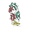
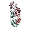
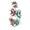
- PDB-7e9b: Structural basis of HLX10 PD-1 receptor recognition, a promising ... -
+ Open data

- Basic information
| Entry | Database: PDB / ID: 7e9b | ||||||
|---|---|---|---|---|---|---|---|
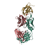
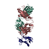
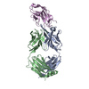
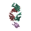
| Title | Structural basis of HLX10 PD-1 receptor recognition, a promising anti-PD-1 antibody clinical candidate for cancer immunotherapy | ||||||
 Components Components |
| ||||||
 Keywords Keywords | IMMUNE SYSTEM / Antibody / mAb / PD-1 / PD-L-1 / Cancer / T-cell / Immunotherapy / ICI | ||||||
| Function / homology |  Function and homology information Function and homology informationnegative regulation of tolerance induction / regulatory T cell apoptotic process / negative regulation of immune response / negative regulation of T cell mediated immune response to tumor cell / negative regulation of T cell activation / positive regulation of T cell apoptotic process / B cell apoptotic process / negative regulation of B cell apoptotic process / humoral immune response / Co-inhibition by PD-1 ...negative regulation of tolerance induction / regulatory T cell apoptotic process / negative regulation of immune response / negative regulation of T cell mediated immune response to tumor cell / negative regulation of T cell activation / positive regulation of T cell apoptotic process / B cell apoptotic process / negative regulation of B cell apoptotic process / humoral immune response / Co-inhibition by PD-1 / regulation of immune response / signaling receptor activity / Potential therapeutics for SARS / adaptive immune response / external side of plasma membrane / apoptotic process / plasma membrane Similarity search - Function | ||||||
| Biological species |  Homo sapiens (human) Homo sapiens (human) | ||||||
| Method |  X-RAY DIFFRACTION / SYNCHROTRON / MOLECULAR REPLACEMENT / molecular replacement / Resolution: 1.78 Å X-RAY DIFFRACTION / SYNCHROTRON / MOLECULAR REPLACEMENT / molecular replacement / Resolution: 1.78 Å | ||||||
 Authors Authors | Fan, S.L. / Jiang, W.D. | ||||||
 Citation Citation | Journal: Plos One / Year: 2021 Title: Structural basis of HLX10 PD-1 receptor recognition, a promising anti-PD-1 antibody clinical candidate for cancer immunotherapy. Authors: Issafras, H. / Fan, S. / Tseng, C.L. / Cheng, Y. / Lin, P. / Xiao, L. / Huang, Y.J. / Tu, C.H. / Hsiao, Y.C. / Li, M. / Chen, Y.H. / Ho, C.H. / Li, O. / Wang, Y. / Chen, S. / Ji, Z. / Zhang, ...Authors: Issafras, H. / Fan, S. / Tseng, C.L. / Cheng, Y. / Lin, P. / Xiao, L. / Huang, Y.J. / Tu, C.H. / Hsiao, Y.C. / Li, M. / Chen, Y.H. / Ho, C.H. / Li, O. / Wang, Y. / Chen, S. / Ji, Z. / Zhang, E. / Mao, Y.T. / Liu, E. / Yang, S. / Jiang, W. | ||||||
| History |
|
- Structure visualization
| Structure viewer | Molecule: MolmilJmol/JSmol |
|---|
- Downloads & links
-Download
| PDBx/mmCIF format | 7e9b.cif.gz | 231.2 KB | Display | PDBx/mmCIF format |
|---|---|---|---|---|
| PDB format | pdb7e9b.ent.gz | 182.7 KB | Display | PDB format |
| PDBx/mmJSON format | 7e9b.json.gz | Tree view | PDBx/mmJSON format | |
| Others |  Other downloads Other downloads |
-Validation report
| Summary document | 7e9b_validation.pdf.gz | 438.5 KB | Display | wwPDB validaton report |
|---|---|---|---|---|
| Full document | 7e9b_full_validation.pdf.gz | 440.9 KB | Display | |
| Data in XML | 7e9b_validation.xml.gz | 25.7 KB | Display | |
| Data in CIF | 7e9b_validation.cif.gz | 37.9 KB | Display | |
| Arichive directory | https://data.pdbj.org/pub/pdb/validation_reports/e9/7e9bftp://data.pdbj.org/pub/pdb/validation_reports/e9/7e9b | HTTPS FTP |
-Related structure data
| Related structure data | 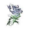 4zqkS S: Starting model for refinement |
|---|---|
| Similar structure data |


-Links
 PDBj
PDBj


- Assembly
| Deposited unit | 
| ||||||||
|---|---|---|---|---|---|---|---|---|---|
| 1 |
| ||||||||
| Unit cell |
|
-Components
| #1: Antibody | Mass: 22834.482 Da / Num. of mol.: 1 Source method: isolated from a genetically manipulated source Source: (gene. exp.) Homo sapiens (human) / Production host:   Cricetulus griseus (Chinese hamster) Cricetulus griseus (Chinese hamster) |
|---|---|
| #2: Antibody | Mass: 23455.074 Da / Num. of mol.: 1 Source method: isolated from a genetically manipulated source Source: (gene. exp.) Homo sapiens (human) / Production host: Cricetulus griseus (Chinese hamster) |
| #3: Protein | Mass: 12916.412 Da / Num. of mol.: 1 Source method: isolated from a genetically manipulated source Source: (gene. exp.) Homo sapiens (human) / Gene: PDCD1, PD1 / Production host:  Escherichia phage EcSzw-2 (virus) / References: UniProt: Q15116 Escherichia phage EcSzw-2 (virus) / References: UniProt: Q15116 |
| #4: Water | ChemComp-HOH /  Mass: 18.015 Da / Num. of mol.: 489 / Source method: isolated from a natural source / Formula: H2O Mass: 18.015 Da / Num. of mol.: 489 / Source method: isolated from a natural source / Formula: H2O |
| Has protein modification | Y |
-Experimental details
-Experiment
| Experiment | Method: X-RAY DIFFRACTION / Number of used crystals: 1 |
|---|
- Sample preparation
| Crystal | Density Matthews: 2.31 Å3/Da / Density % sol: 46.86 % |
|---|---|
| Crystal grow | Temperature: 291 K / Method: vapor diffusion, sitting drop / Details: 0.1 M Tris pH 7.5 containing 23% (w/v) PEG 4000 |
-Data collection
| Diffraction | Mean temperature: 100 K / Serial crystal experiment: N |
|---|---|
| Diffraction source | Source: SYNCHROTRON / Site: SSRF  / Beamline: BL19U1 / Wavelength: 0.9751 Å / Beamline: BL19U1 / Wavelength: 0.9751 Å |
| Detector | Type: DECTRIS PILATUS 6M / Detector: PIXEL / Date: Oct 21, 2020 |
| Radiation | Protocol: SINGLE WAVELENGTH / Monochromatic (M) / Laue (L): M / Scattering type: x-ray |
| Radiation wavelength | Wavelength: 0.9751 Å / Relative weight: 1 |
| Reflection | Resolution: 1.779→31.414 Å / Num. obs: 48174 / % possible obs: 94.63 % / Redundancy: 7.1 % / CC1/2: 0.989 / Net I/σ(I): 28.9 |
| Reflection shell | Resolution: 1.779→1.84 Å / Num. unique obs: 48174 / CC1/2: 0.989 |
-Phasing
| Phasing | Method: molecular replacement |
|---|
- Processing
| Software |
| ||||||||||||||||||||||||||||||||||||||||||||||||||||||||||||||||||||||||||||||||||||||||||||||||||||||||||||||||||||||||||||||||||||||||||||||||||||||||||||||||||||||||||||||||||||||||||||||||||||||||||||||||||||||||||||||||||||||||||||||||||||||||||||||||||||||||||||||||||||||||||||||||||||||||||||||||||||||||||||||||||||||||||||||||||||||||||||||
|---|---|---|---|---|---|---|---|---|---|---|---|---|---|---|---|---|---|---|---|---|---|---|---|---|---|---|---|---|---|---|---|---|---|---|---|---|---|---|---|---|---|---|---|---|---|---|---|---|---|---|---|---|---|---|---|---|---|---|---|---|---|---|---|---|---|---|---|---|---|---|---|---|---|---|---|---|---|---|---|---|---|---|---|---|---|---|---|---|---|---|---|---|---|---|---|---|---|---|---|---|---|---|---|---|---|---|---|---|---|---|---|---|---|---|---|---|---|---|---|---|---|---|---|---|---|---|---|---|---|---|---|---|---|---|---|---|---|---|---|---|---|---|---|---|---|---|---|---|---|---|---|---|---|---|---|---|---|---|---|---|---|---|---|---|---|---|---|---|---|---|---|---|---|---|---|---|---|---|---|---|---|---|---|---|---|---|---|---|---|---|---|---|---|---|---|---|---|---|---|---|---|---|---|---|---|---|---|---|---|---|---|---|---|---|---|---|---|---|---|---|---|---|---|---|---|---|---|---|---|---|---|---|---|---|---|---|---|---|---|---|---|---|---|---|---|---|---|---|---|---|---|---|---|---|---|---|---|---|---|---|---|---|---|---|---|---|---|---|---|---|---|---|---|---|---|---|---|---|---|---|---|---|---|---|---|---|---|---|---|---|---|---|---|---|---|---|---|---|---|---|---|---|---|---|---|---|---|---|---|---|---|---|---|---|---|---|---|---|---|---|---|---|---|---|---|---|---|---|---|---|---|---|---|---|---|---|---|---|---|---|---|---|---|---|---|---|---|---|---|---|---|
| Refinement | Method to determine structure: MOLECULAR REPLACEMENT Starting model: 4ZQK Resolution: 1.78→31.41 Å / SU ML: 0.18 / Cross valid method: THROUGHOUT / σ(F): 1.97 / Phase error: 22.27 / Stereochemistry target values: ML
| ||||||||||||||||||||||||||||||||||||||||||||||||||||||||||||||||||||||||||||||||||||||||||||||||||||||||||||||||||||||||||||||||||||||||||||||||||||||||||||||||||||||||||||||||||||||||||||||||||||||||||||||||||||||||||||||||||||||||||||||||||||||||||||||||||||||||||||||||||||||||||||||||||||||||||||||||||||||||||||||||||||||||||||||||||||||||||||||
| Solvent computation | Shrinkage radii: 0.9 Å / VDW probe radii: 1.11 Å / Solvent model: FLAT BULK SOLVENT MODEL | ||||||||||||||||||||||||||||||||||||||||||||||||||||||||||||||||||||||||||||||||||||||||||||||||||||||||||||||||||||||||||||||||||||||||||||||||||||||||||||||||||||||||||||||||||||||||||||||||||||||||||||||||||||||||||||||||||||||||||||||||||||||||||||||||||||||||||||||||||||||||||||||||||||||||||||||||||||||||||||||||||||||||||||||||||||||||||||||
| Displacement parameters | Biso max: 88.89 Å2 / Biso mean: 32.7236 Å2 / Biso min: 14.05 Å2 | ||||||||||||||||||||||||||||||||||||||||||||||||||||||||||||||||||||||||||||||||||||||||||||||||||||||||||||||||||||||||||||||||||||||||||||||||||||||||||||||||||||||||||||||||||||||||||||||||||||||||||||||||||||||||||||||||||||||||||||||||||||||||||||||||||||||||||||||||||||||||||||||||||||||||||||||||||||||||||||||||||||||||||||||||||||||||||||||
| Refinement step | Cycle: final / Resolution: 1.78→31.41 Å
| ||||||||||||||||||||||||||||||||||||||||||||||||||||||||||||||||||||||||||||||||||||||||||||||||||||||||||||||||||||||||||||||||||||||||||||||||||||||||||||||||||||||||||||||||||||||||||||||||||||||||||||||||||||||||||||||||||||||||||||||||||||||||||||||||||||||||||||||||||||||||||||||||||||||||||||||||||||||||||||||||||||||||||||||||||||||||||||||
| LS refinement shell | Refine-ID: X-RAY DIFFRACTION / Rfactor Rfree error: 0 / Total num. of bins used: 14
| ||||||||||||||||||||||||||||||||||||||||||||||||||||||||||||||||||||||||||||||||||||||||||||||||||||||||||||||||||||||||||||||||||||||||||||||||||||||||||||||||||||||||||||||||||||||||||||||||||||||||||||||||||||||||||||||||||||||||||||||||||||||||||||||||||||||||||||||||||||||||||||||||||||||||||||||||||||||||||||||||||||||||||||||||||||||||||||||
| Refinement TLS params. | Method: refined / Refine-ID: X-RAY DIFFRACTION
| ||||||||||||||||||||||||||||||||||||||||||||||||||||||||||||||||||||||||||||||||||||||||||||||||||||||||||||||||||||||||||||||||||||||||||||||||||||||||||||||||||||||||||||||||||||||||||||||||||||||||||||||||||||||||||||||||||||||||||||||||||||||||||||||||||||||||||||||||||||||||||||||||||||||||||||||||||||||||||||||||||||||||||||||||||||||||||||||
| Refinement TLS group |
|