 Movie
Movie Controller

[English] 日本語
 Yorodumi
- PDB-7e6g: Crystal structure of diguanylate cyclase SiaD in complex with its... -
+ Open data

- Basic information
| Entry | Database: PDB / ID: 7e6g | |||||||||
|---|---|---|---|---|---|---|---|---|---|---|
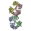
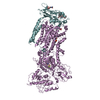
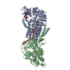
| Title | Crystal structure of diguanylate cyclase SiaD in complex with its activator SiaC from Pseudomonas aeruginosa | |||||||||
 Components Components |
| |||||||||
 Keywords Keywords | DE NOVO PROTEIN / diguanylate cyclase / activation / Pseudomonas aeruginosa / BIOSYNTHETIC PROTEIN | |||||||||
| Function / homology |  Function and homology information Function and homology information | |||||||||
| Biological species |   Pseudomonas aeruginosa (bacteria) Pseudomonas aeruginosa (bacteria) | |||||||||
| Method |  X-RAY DIFFRACTION / SYNCHROTRON / MOLECULAR REPLACEMENT / Resolution: 2.65 Å X-RAY DIFFRACTION / SYNCHROTRON / MOLECULAR REPLACEMENT / Resolution: 2.65 Å | |||||||||
 Authors Authors | Zhou, J.S. / Zhang, L. / Zhang, L. | |||||||||
| Funding support |  China, 2items China, 2items
| |||||||||
 Citation Citation | Journal: Elife / Year: 2021 Title: Structural basis for diguanylate cyclase activation by its binding partner in Pseudomonas aeruginosa . Authors: Chen, G. / Zhou, J. / Zuo, Y. / Huo, W. / Peng, J. / Li, M. / Zhang, Y. / Wang, T. / Zhang, L. / Zhang, L. / Liang, H. | |||||||||
| History |
|
- Structure visualization
| Structure viewer | Molecule: MolmilJmol/JSmol |
|---|
- Downloads & links
-Download
| PDBx/mmCIF format | 7e6g.cif.gz | 222 KB | Display | PDBx/mmCIF format |
|---|---|---|---|---|
| PDB format | pdb7e6g.ent.gz | 175.1 KB | Display | PDB format |
| PDBx/mmJSON format | 7e6g.json.gz | Tree view | PDBx/mmJSON format | |
| Others |  Other downloads Other downloads |
-Validation report
| Summary document | 7e6g_validation.pdf.gz | 1.1 MB | Display | wwPDB validaton report |
|---|---|---|---|---|
| Full document | 7e6g_full_validation.pdf.gz | 1.1 MB | Display | |
| Data in XML | 7e6g_validation.xml.gz | 42.3 KB | Display | |
| Data in CIF | 7e6g_validation.cif.gz | 58.2 KB | Display | |
| Arichive directory | https://data.pdbj.org/pub/pdb/validation_reports/e6/7e6gftp://data.pdbj.org/pub/pdb/validation_reports/e6/7e6g | HTTPS FTP |
-Related structure data
| Related structure data |  3i5aS S: Starting model for refinement |
|---|---|
| Similar structure data |


-Links
 PDBj
PDBj


- Assembly
| Deposited unit | 
| ||||||||
|---|---|---|---|---|---|---|---|---|---|
| 1 |
| ||||||||
| Unit cell |
|
-Components
| #1: Protein | Mass: 31587.918 Da / Num. of mol.: 2 Source method: isolated from a genetically manipulated source Source: (gene. exp.) Pseudomonas aeruginosa (bacteria)Gene: pleD_3, pleD_1, pleD_4, NCTC12951_03415, NCTC13437_04900, NCTC13621_04785, PAMH19_0165, RW109_RW109_00771 Production host: #2: Protein | Mass: 14655.237 Da / Num. of mol.: 4 Source method: isolated from a genetically manipulated source Source: (gene. exp.) Pseudomonas aeruginosa (bacteria) / Production host: #3: Chemical | ChemComp-MG / |   Mass: 24.305 Da / Num. of mol.: 1 / Source method: obtained synthetically / Formula: Mg / Feature type: SUBJECT OF INVESTIGATION Mass: 24.305 Da / Num. of mol.: 1 / Source method: obtained synthetically / Formula: Mg / Feature type: SUBJECT OF INVESTIGATION#4: Chemical | ChemComp-G2P / |   Mass: 521.208 Da / Num. of mol.: 1 / Source method: obtained synthetically / Formula: C11H18N5O13P3 / Feature type: SUBJECT OF INVESTIGATION / Comment: GMP-CPP, energy-carrying molecule analogue*YM Mass: 521.208 Da / Num. of mol.: 1 / Source method: obtained synthetically / Formula: C11H18N5O13P3 / Feature type: SUBJECT OF INVESTIGATION / Comment: GMP-CPP, energy-carrying molecule analogue*YM#5: Water | ChemComp-HOH / |  Mass: 18.015 Da / Num. of mol.: 197 / Source method: isolated from a natural source / Formula: H2O Mass: 18.015 Da / Num. of mol.: 197 / Source method: isolated from a natural source / Formula: H2OHas ligand of interest | Y | |
|---|
-Experimental details
-Experiment
| Experiment | Method: X-RAY DIFFRACTION / Number of used crystals: 1 |
|---|
- Sample preparation
| Crystal | Density Matthews: 2.98 Å3/Da / Density % sol: 58.72 % |
|---|---|
| Crystal grow | Temperature: 277 K / Method: vapor diffusion, sitting drop Details: 0.01 M Spermidine trihydrochloride and 15% w/v PEG 3350 |
-Data collection
| Diffraction | Mean temperature: 100 K / Serial crystal experiment: N |
|---|---|
| Diffraction source | Source: SYNCHROTRON / Site: SSRF / Beamline: BL19U1 / Wavelength: 0.9785 Å |
| Detector | Type: DECTRIS PILATUS3 6M / Detector: PIXEL / Date: Jan 2, 2019 |
| Radiation | Protocol: SINGLE WAVELENGTH / Monochromatic (M) / Laue (L): M / Scattering type: x-ray |
| Radiation wavelength | Wavelength: 0.9785 Å / Relative weight: 1 |
| Reflection | Resolution: 2.65→50 Å / Num. obs: 40897 / % possible obs: 99.5 % / Redundancy: 13.3 % / CC1/2: 0.982 / Net I/σ(I): 13.1 |
| Reflection shell | Resolution: 2.65→2.74 Å / Num. unique obs: 4204 / CC1/2: 0.588 |
- Processing
| Software |
| ||||||||||||||||||||||||||||||||||||||||||||||||||||||||||||||||||||||||||||||||||||||||||||||||
|---|---|---|---|---|---|---|---|---|---|---|---|---|---|---|---|---|---|---|---|---|---|---|---|---|---|---|---|---|---|---|---|---|---|---|---|---|---|---|---|---|---|---|---|---|---|---|---|---|---|---|---|---|---|---|---|---|---|---|---|---|---|---|---|---|---|---|---|---|---|---|---|---|---|---|---|---|---|---|---|---|---|---|---|---|---|---|---|---|---|---|---|---|---|---|---|---|---|
| Refinement | Method to determine structure: MOLECULAR REPLACEMENT Starting model: 3I5A Resolution: 2.65→46.295 Å / SU ML: 0.36 / Cross valid method: THROUGHOUT / σ(F): 1.34 / Phase error: 27.71 / Stereochemistry target values: ML
| ||||||||||||||||||||||||||||||||||||||||||||||||||||||||||||||||||||||||||||||||||||||||||||||||
| Solvent computation | Shrinkage radii: 0.9 Å / VDW probe radii: 1.11 Å / Solvent model: FLAT BULK SOLVENT MODEL | ||||||||||||||||||||||||||||||||||||||||||||||||||||||||||||||||||||||||||||||||||||||||||||||||
| Displacement parameters | Biso max: 132.86 Å2 / Biso mean: 55.2951 Å2 / Biso min: 11.88 Å2 | ||||||||||||||||||||||||||||||||||||||||||||||||||||||||||||||||||||||||||||||||||||||||||||||||
| Refinement step | Cycle: final / Resolution: 2.65→46.295 Å
| ||||||||||||||||||||||||||||||||||||||||||||||||||||||||||||||||||||||||||||||||||||||||||||||||
| LS refinement shell | Refine-ID: X-RAY DIFFRACTION / Rfactor Rfree error: 0
|
