 Movie
Movie Controller


+ Open data

- Basic information
| Entry | Database: PDB / ID: 7.0E+57 | ||||||
|---|---|---|---|---|---|---|---|
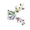
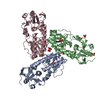
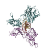
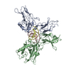
| Title | Crystal structure of murine GITR-GITRL complex | ||||||
 Components Components |
| ||||||
 Keywords Keywords | IMMUNE SYSTEM / Complex | ||||||
| Function / homology |  Function and homology information Function and homology informationregulation of dendritic cell chemotaxis / TNFs bind their physiological receptors / tumor necrosis factor receptor activity / tumor necrosis factor receptor superfamily binding / negative regulation of T-helper 17 cell lineage commitment / tumor necrosis factor receptor binding / positive regulation of monocyte chemotaxis / T cell proliferation involved in immune response / positive regulation of leukocyte migration / positive regulation of macrophage chemotaxis ...regulation of dendritic cell chemotaxis / TNFs bind their physiological receptors / tumor necrosis factor receptor activity / tumor necrosis factor receptor superfamily binding / negative regulation of T-helper 17 cell lineage commitment / tumor necrosis factor receptor binding / positive regulation of monocyte chemotaxis / T cell proliferation involved in immune response / positive regulation of leukocyte migration / positive regulation of macrophage chemotaxis / regulation of T cell proliferation / regulation of protein-containing complex assembly / positive regulation of cell adhesion / cytokine activity / tumor necrosis factor-mediated signaling pathway / : / positive regulation of inflammatory response / adaptive immune response / external side of plasma membrane / apoptotic process / negative regulation of apoptotic process / cell surface / extracellular space / extracellular region / identical protein binding / plasma membrane Similarity search - Function | ||||||
| Biological species |  | ||||||
| Method |  X-RAY DIFFRACTION / SYNCHROTRON / MOLECULAR REPLACEMENT / Resolution: 3.302 Å X-RAY DIFFRACTION / SYNCHROTRON / MOLECULAR REPLACEMENT / Resolution: 3.302 Å | ||||||
 Authors Authors | Zhao, M. / Tan, S. / Fu, L. / Chai, Y. / Qi, J. / Gao, G.F. | ||||||
 Citation Citation | Journal: Cell Rep / Year: 2021 Title: Atypical TNF-TNFR superfamily binding interface in the GITR-GITRL complex for T cell activation. Authors: Zhao, M. / Fu, L. / Chai, Y. / Sun, M. / Li, Y. / Wang, S. / Qi, J. / Zeng, B. / Kang, L. / Gao, G.F. / Tan, S. | ||||||
| History |
|
- Structure visualization
| Structure viewer | Molecule: MolmilJmol/JSmol |
|---|
- Downloads & links
-Download
| PDBx/mmCIF format | 7e57.cif.gz | 108.6 KB | Display | PDBx/mmCIF format |
|---|---|---|---|---|
| PDB format | pdb7e57.ent.gz | 79.5 KB | Display | PDB format |
| PDBx/mmJSON format | 7e57.json.gz | Tree view | PDBx/mmJSON format | |
| Others |  Other downloads Other downloads |
-Validation report
| Arichive directory | https://data.pdbj.org/pub/pdb/validation_reports/e5/7e57ftp://data.pdbj.org/pub/pdb/validation_reports/e5/7e57 | HTTPS FTP |
|---|
-Related structure data


-Links
 PDBj
PDBj
- Assembly
| Deposited unit | 
| ||||||||
|---|---|---|---|---|---|---|---|---|---|
| 1 |
| ||||||||
| Unit cell |
|
-Components
| #1: Protein | Mass: 19755.266 Da / Num. of mol.: 2 Source method: isolated from a genetically manipulated source Source: (gene. exp.)  #2: Protein | Mass: 25360.127 Da / Num. of mol.: 2 Source method: isolated from a genetically manipulated source Source: (gene. exp.) Production host: Insect cell expression vector pTIE1 (others) References: UniProt: O35714 #3: Polysaccharide | alpha-L-fucopyranose-(1-6)-2-acetamido-2-deoxy-beta-D-glucopyranose | Source method: isolated from a genetically manipulated source #4: Polysaccharide | 2-acetamido-2-deoxy-beta-D-glucopyranose-(1-4)-[alpha-L-fucopyranose-(1-6)]2-acetamido-2-deoxy-beta- ...2-acetamido-2-deoxy-beta-D-glucopyranose-(1-4)-[alpha-L-fucopyranose-(1-6)]2-acetamido-2-deoxy-beta-D-glucopyranose | Source method: isolated from a genetically manipulated source Has ligand of interest | N | Has protein modification | Y | |
|---|
-Experimental details
-Experiment
| Experiment | Method: X-RAY DIFFRACTION / Number of used crystals: 1 |
|---|
- Sample preparation
| Crystal | Density Matthews: 2.14 Å3/Da / Density % sol: 42.63 % |
|---|---|
| Crystal grow | Temperature: 277 K / Method: vapor diffusion, sitting drop Details: 0.02M nickel (II) chloride hexahydrate, 0.02M magnesium chloride hexahydrate, 0.02M cadmium chloride hydrate, 0.1M sodium acetate trihydrate, pH4.1, 20% w/v polyethylene glycol monomethyl ether 2000 |
-Data collection
| Diffraction | Mean temperature: 100 K / Serial crystal experiment: N |
|---|---|
| Diffraction source | Source: SYNCHROTRON / Site: SSRF  / Beamline: BL17U1 / Wavelength: 1.03923 Å / Beamline: BL17U1 / Wavelength: 1.03923 Å |
| Detector | Type: ADSC QUANTUM 315r / Detector: CCD / Date: Apr 7, 2019 |
| Radiation | Protocol: SINGLE WAVELENGTH / Monochromatic (M) / Laue (L): M / Scattering type: x-ray |
| Radiation wavelength | Wavelength: 1.03923 Å / Relative weight: 1 |
| Reflection | Resolution: 3.3→50 Å / Num. obs: 77449 / % possible obs: 84.98 % / Redundancy: 6.9 % / CC1/2: 0.856 / Rmerge(I) obs: 0.094 / Net I/σ(I): 19.29 |
| Reflection shell | Resolution: 3.3→3.42 Å / Rmerge(I) obs: 0.778 / CC1/2: 0.848 |
- Processing
| Software |
| |||||||||||||||||||||||||||||||||||
|---|---|---|---|---|---|---|---|---|---|---|---|---|---|---|---|---|---|---|---|---|---|---|---|---|---|---|---|---|---|---|---|---|---|---|---|---|
| Refinement | Method to determine structure: MOLECULAR REPLACEMENT Starting model: 2Q8O, 6A3V Resolution: 3.302→38.463 Å / SU ML: 0.5 / Cross valid method: FREE R-VALUE / σ(F): 1.37 / Phase error: 34.83 / Stereochemistry target values: ML
| |||||||||||||||||||||||||||||||||||
| Solvent computation | Shrinkage radii: 0.9 Å / VDW probe radii: 1.11 Å / Solvent model: FLAT BULK SOLVENT MODEL | |||||||||||||||||||||||||||||||||||
| Refinement step | Cycle: LAST / Resolution: 3.302→38.463 Å
| |||||||||||||||||||||||||||||||||||
| Refine LS restraints |
| |||||||||||||||||||||||||||||||||||
| LS refinement shell |
|
