 Movie
Movie Controller

[English] 日本語
 Yorodumi
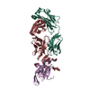
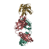
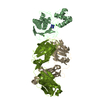
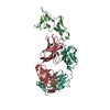
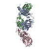
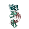
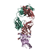
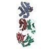
- PDB-7e3o: Crystal structure of SARS-CoV-2 receptor binding domain in comple... -
+ Open data

- Basic information
| Entry | Database: PDB / ID: 7e3o | ||||||
|---|---|---|---|---|---|---|---|
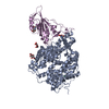
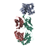
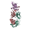
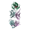
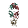
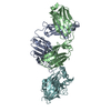
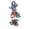
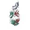
| Title | Crystal structure of SARS-CoV-2 receptor binding domain in complex with neutralizing antibody nCoV617 | ||||||
 Components Components |
| ||||||
 Keywords Keywords | VIRAL PROTEIN / SARS-CoV-2 / S-RBD / RBS-C / native human monoclonal antibody / therapeutic antibody | ||||||
| Function / homology |  Function and homology information Function and homology informationsymbiont-mediated disruption of host tissue / Maturation of spike protein / Translation of Structural Proteins / Virion Assembly and Release / host cell surface / host extracellular space / viral translation / symbiont-mediated-mediated suppression of host tetherin activity / Induction of Cell-Cell Fusion / structural constituent of virion ...symbiont-mediated disruption of host tissue / Maturation of spike protein / Translation of Structural Proteins / Virion Assembly and Release / host cell surface / host extracellular space / viral translation / symbiont-mediated-mediated suppression of host tetherin activity / Induction of Cell-Cell Fusion / structural constituent of virion / membrane fusion / entry receptor-mediated virion attachment to host cell / Attachment and Entry / host cell endoplasmic reticulum-Golgi intermediate compartment membrane / positive regulation of viral entry into host cell / receptor-mediated virion attachment to host cell / host cell surface receptor binding / symbiont-mediated suppression of host innate immune response / receptor ligand activity / endocytosis involved in viral entry into host cell / fusion of virus membrane with host plasma membrane / fusion of virus membrane with host endosome membrane / viral envelope / symbiont entry into host cell / virion attachment to host cell / SARS-CoV-2 activates/modulates innate and adaptive immune responses / host cell plasma membrane / virion membrane / identical protein binding / membrane / plasma membrane Similarity search - Function | ||||||
| Biological species |  Homo sapiens (human) Homo sapiens (human)  Severe acute respiratory syndrome coronavirus 2 Severe acute respiratory syndrome coronavirus 2 | ||||||
| Method |  X-RAY DIFFRACTION / SYNCHROTRON / MOLECULAR REPLACEMENT / Resolution: 2.51 Å X-RAY DIFFRACTION / SYNCHROTRON / MOLECULAR REPLACEMENT / Resolution: 2.51 Å | ||||||
 Authors Authors | Chen, S.D. / Yang, M. | ||||||
 Citation Citation | Journal: Microbiol Spectr / Year: 2021 Title: Structural Basis of a Human Neutralizing Antibody Specific to the SARS-CoV-2 Spike Protein Receptor-Binding Domain. Authors: Yang, M. / Li, J. / Huang, Z. / Li, H. / Wang, Y. / Wang, X. / Kang, S. / Huang, X. / Wu, C. / Liu, T. / Jia, Z. / Liang, J. / Yuan, X. / He, S. / Chen, X. / Zhou, Z. / Chen, Q. / Liu, S. / ...Authors: Yang, M. / Li, J. / Huang, Z. / Li, H. / Wang, Y. / Wang, X. / Kang, S. / Huang, X. / Wu, C. / Liu, T. / Jia, Z. / Liang, J. / Yuan, X. / He, S. / Chen, X. / Zhou, Z. / Chen, Q. / Liu, S. / Li, J. / Zheng, H. / Liu, X. / Li, K. / Yao, X. / Lang, B. / Liu, L. / Liao, H.X. / Chen, S. | ||||||
| History |
|
- Structure visualization
| Structure viewer | Molecule: MolmilJmol/JSmol |
|---|
- Downloads & links
-Download
| PDBx/mmCIF format | 7e3o.cif.gz | 292.5 KB | Display | PDBx/mmCIF format |
|---|---|---|---|---|
| PDB format | pdb7e3o.ent.gz | 195.1 KB | Display | PDB format |
| PDBx/mmJSON format | 7e3o.json.gz | Tree view | PDBx/mmJSON format | |
| Others |  Other downloads Other downloads |
-Validation report
| Arichive directory | https://data.pdbj.org/pub/pdb/validation_reports/e3/7e3oftp://data.pdbj.org/pub/pdb/validation_reports/e3/7e3o | HTTPS FTP |
|---|
-Related structure data


-Links
 PDBj
PDBj


- Assembly
| Deposited unit | 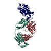
| ||||||||||||
|---|---|---|---|---|---|---|---|---|---|---|---|---|---|
| 1 |
| ||||||||||||
| Unit cell |
|
-Components
| #1: Antibody | Mass: 22551.846 Da / Num. of mol.: 1 Source method: isolated from a genetically manipulated source Source: (gene. exp.) Homo sapiens (human)Production host: Mammalian expression vector HA-MCS-pcDNA3.1 (others) |
|---|---|
| #2: Antibody | Mass: 24278.334 Da / Num. of mol.: 1 Source method: isolated from a genetically manipulated source Source: (gene. exp.) Homo sapiens (human)Production host: Mammalian expression vector HA-MCS-pcDNA3.1 (others) |
| #3: Protein | Mass: 22547.164 Da / Num. of mol.: 1 / Fragment: UNP residues 337-527 Source method: isolated from a genetically manipulated source Source: (gene. exp.) Severe acute respiratory syndrome coronavirus 2Gene: S, 2 / Production host: unidentified baculovirus / References: UniProt: P0DTC2 |
| #4: Water | ChemComp-HOH /  Mass: 18.015 Da / Num. of mol.: 63 / Source method: isolated from a natural source / Formula: H2O Mass: 18.015 Da / Num. of mol.: 63 / Source method: isolated from a natural source / Formula: H2O |
| Has protein modification | Y |
-Experimental details
-Experiment
| Experiment | Method: X-RAY DIFFRACTION / Number of used crystals: 1 |
|---|
- Sample preparation
| Crystal | Density Matthews: 2.78 Å3/Da / Density % sol: 55.69 % |
|---|---|
| Crystal grow | Temperature: 289.15 K / Method: vapor diffusion, hanging drop / Details: 152 mM NH4Cl, 22.8% PEG8000 |
-Data collection
| Diffraction | Mean temperature: 100 K / Serial crystal experiment: N |
|---|---|
| Diffraction source | Source: SYNCHROTRON / Site: SSRF  / Beamline: BL19U1 / Wavelength: 0.97853 Å / Beamline: BL19U1 / Wavelength: 0.97853 Å |
| Detector | Type: SDMS / Detector: CMOS / Date: Nov 27, 2020 |
| Radiation | Protocol: SINGLE WAVELENGTH / Monochromatic (M) / Laue (L): M / Scattering type: x-ray |
| Radiation wavelength | Wavelength: 0.97853 Å / Relative weight: 1 |
| Reflection | Resolution: 2.51→40.67 Å / Num. obs: 25491 / % possible obs: 94.2 % / Redundancy: 4.9 % / Biso Wilson estimate: 54 Å2 / CC1/2: 0.897 / Net I/σ(I): -0.6 |
| Reflection shell | Resolution: 2.514→2.603 Å / Num. unique obs: 2495 / CC1/2: 0.868 |
- Processing
| Software |
| |||||||||||||||||||||||||||||||||||||||||||||||||||||||||||||||||||||||||||||||||||||||||||||||||||||||||
|---|---|---|---|---|---|---|---|---|---|---|---|---|---|---|---|---|---|---|---|---|---|---|---|---|---|---|---|---|---|---|---|---|---|---|---|---|---|---|---|---|---|---|---|---|---|---|---|---|---|---|---|---|---|---|---|---|---|---|---|---|---|---|---|---|---|---|---|---|---|---|---|---|---|---|---|---|---|---|---|---|---|---|---|---|---|---|---|---|---|---|---|---|---|---|---|---|---|---|---|---|---|---|---|---|---|---|
| Refinement | Method to determine structure: MOLECULAR REPLACEMENT Starting model: 6M0J, 7K8Z Resolution: 2.51→40.67 Å / SU ML: 0.3431 / Cross valid method: FREE R-VALUE / σ(F): 1.36 / Phase error: 29.0598 Stereochemistry target values: GeoStd + Monomer Library + CDL v1.2
| |||||||||||||||||||||||||||||||||||||||||||||||||||||||||||||||||||||||||||||||||||||||||||||||||||||||||
| Solvent computation | Shrinkage radii: 0.9 Å / VDW probe radii: 1.11 Å / Solvent model: FLAT BULK SOLVENT MODEL | |||||||||||||||||||||||||||||||||||||||||||||||||||||||||||||||||||||||||||||||||||||||||||||||||||||||||
| Displacement parameters | Biso mean: 55.17 Å2 | |||||||||||||||||||||||||||||||||||||||||||||||||||||||||||||||||||||||||||||||||||||||||||||||||||||||||
| Refinement step | Cycle: LAST / Resolution: 2.51→40.67 Å
| |||||||||||||||||||||||||||||||||||||||||||||||||||||||||||||||||||||||||||||||||||||||||||||||||||||||||
| Refine LS restraints |
| |||||||||||||||||||||||||||||||||||||||||||||||||||||||||||||||||||||||||||||||||||||||||||||||||||||||||
| LS refinement shell |
| |||||||||||||||||||||||||||||||||||||||||||||||||||||||||||||||||||||||||||||||||||||||||||||||||||||||||
| Refinement TLS params. | Method: refined / Origin x: -17.2087125013 Å / Origin y: 8.26329743651 Å / Origin z: -6.48148426029 Å
| |||||||||||||||||||||||||||||||||||||||||||||||||||||||||||||||||||||||||||||||||||||||||||||||||||||||||
| Refinement TLS group | Selection details: all |
