 Movie
Movie Controller

[English] 日本語
 Yorodumi
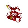
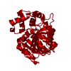
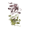
- PDB-7e00: Trans-proline-hydroxylase H11 with Succinic and L-proline in the ... -
+ Open data

- Basic information
| Entry | Database: PDB / ID: 7 | ||||||
|---|---|---|---|---|---|---|---|
| Title | Trans-proline-hydroxylase H11 with Succinic and L-proline in the fourth reaction state. | ||||||
 Components Components | Phytanoyl-CoA dioxygenase | ||||||
 Keywords Keywords | HYDROLASE / L-proline / Trans / Hydroxylase / AKG / mutant | ||||||
| Function / homology | Phytanoyl-CoA dioxygenase / Phytanoyl-CoA dioxygenase (PhyH) / 2-oxoglutarate-dependent dioxygenase activity / iron ion binding / 2-OXOGLUTARIC ACID / OXYGEN ATOM / PROLINE / SUCCINIC ACID / Phytanoyl-CoA dioxygenase Function and homology information Function and homology information | ||||||
| Biological species |  uncultured bacterium esnapd13 (environmental samples) uncultured bacterium esnapd13 (environmental samples) | ||||||
| Method |  X-RAY DIFFRACTION / SYNCHROTRON / MOLECULAR REPLACEMENT / Resolution: 1.92 Å X-RAY DIFFRACTION / SYNCHROTRON / MOLECULAR REPLACEMENT / Resolution: 1.92 Å | ||||||
 Authors Authors | Gong, W.G. / Yang, L.Y. | ||||||
| Funding support |  China, 1items China, 1items
| ||||||
 Citation Citation | Journal: To Be Published Title: Trans-3/4-proline-hydroxylase H11 with AKG and L-proline Authors: Gong, W.G. / Yang, L.Y. | ||||||
| History |
|
- Structure visualization
| Structure viewer | Molecule: MolmilJmol/JSmol |
|---|
- Downloads & links
-Download
| PDBx/mmCIF format | 7e00.cif.gz | 122.4 KB | Display | PDBx/mmCIF format |
|---|---|---|---|---|
| PDB format | pdb7e00.ent.gz | 95.2 KB | Display | PDB format |
| PDBx/mmJSON format | 7e00.json.gz | Tree view | PDBx/mmJSON format | |
| Others |  Other downloads Other downloads |
-Validation report
| Arichive directory | https://data.pdbj.org/pub/pdb/validation_reports/e0/7e00ftp://data.pdbj.org/pub/pdb/validation_reports/e0/7e00 | HTTPS FTP |
|---|
-Related structure data
| Related structure data |  7dt0SC  7e05C  7e06C  7e07C  7e09C S: Starting model for refinement C: citing same article ( |
|---|---|
| Similar structure data |


-Links
 PDBj
PDBj


- Assembly
| Deposited unit | 
| ||||||||
|---|---|---|---|---|---|---|---|---|---|
| 1 |
| ||||||||
| Unit cell |
|
-Components
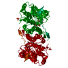
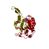
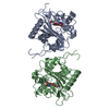
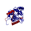
-Protein , 1 types, 2 molecules AB
| #1: Protein | Mass: 30108.789 Da / Num. of mol.: 2 Source method: isolated from a genetically manipulated source Source: (gene. exp.) uncultured bacterium esnapd13 (environmental samples)Production host: |
|---|
-Non-polymers , 5 types, 242 molecules 


| #2: Chemical |  Type: L-peptide linking / Mass: 115.130 Da / Num. of mol.: 2 / Source method: obtained synthetically / Formula: C5H9NO2 / Feature type: SUBJECT OF INVESTIGATION Type: L-peptide linking / Mass: 115.130 Da / Num. of mol.: 2 / Source method: obtained synthetically / Formula: C5H9NO2 / Feature type: SUBJECT OF INVESTIGATION#3: Chemical |  Mass: 118.088 Da / Num. of mol.: 2 / Source method: obtained synthetically / Formula: C4H6O4 / Feature type: SUBJECT OF INVESTIGATION Mass: 118.088 Da / Num. of mol.: 2 / Source method: obtained synthetically / Formula: C4H6O4 / Feature type: SUBJECT OF INVESTIGATION#4: Chemical | ChemComp-AKG / |  Mass: 146.098 Da / Num. of mol.: 1 / Source method: obtained synthetically / Formula: C5H6O5 / Feature type: SUBJECT OF INVESTIGATION Mass: 146.098 Da / Num. of mol.: 1 / Source method: obtained synthetically / Formula: C5H6O5 / Feature type: SUBJECT OF INVESTIGATION#5: Chemical | ChemComp-O / |  Mass: 15.999 Da / Num. of mol.: 1 / Source method: obtained synthetically / Formula: O / Feature type: SUBJECT OF INVESTIGATION Mass: 15.999 Da / Num. of mol.: 1 / Source method: obtained synthetically / Formula: O / Feature type: SUBJECT OF INVESTIGATION#6: Water | ChemComp-HOH / | Mass: 18.015 Da / Num. of mol.: 236 / Source method: isolated from a natural source / Formula: H2O |
|---|
-Details
| Has ligand of interest | Y |
|---|
-Experimental details
-Experiment
| Experiment | Method: X-RAY DIFFRACTION / Number of used crystals: 1 |
|---|
- Sample preparation
| Crystal | Density Matthews: 3.38 Å3/Da / Density % sol: 63.61 % |
|---|---|
| Crystal grow | Temperature: 289.15 K / Method: vapor diffusion, sitting drop / pH: 7 Details: 0.7 M Sodium citrate tribasic dihydrate, 0.1 M Bis-Tris propane (pH 7.0) |
-Data collection
| Diffraction | Mean temperature: 100 K / Serial crystal experiment: N |
|---|---|
| Diffraction source | Source: SYNCHROTRON / Site: SSRF / Beamline: BL17U / Wavelength: 0.979 Å |
| Detector | Type: DECTRIS EIGER X 16M / Detector: PIXEL / Date: Oct 26, 2020 |
| Radiation | Protocol: SINGLE WAVELENGTH / Monochromatic (M) / Laue (L): M / Scattering type: x-ray |
| Radiation wavelength | Wavelength: 0.979 Å / Relative weight: 1 |
| Reflection | Resolution: 1.92→85.54 Å / Num. obs: 63525 / % possible obs: 99.2 % / Redundancy: 13 % / Rmerge(I) obs: 0.136 / Net I/σ(I): 11.2 |
| Reflection shell | Resolution: 1.92→2.02 Å / Rmerge(I) obs: 1.218 / Num. unique obs: 9204 / Rpim(I) all: 0.351 / % possible all: 100 |
- Processing
| Software |
| ||||||||||||||||||||
|---|---|---|---|---|---|---|---|---|---|---|---|---|---|---|---|---|---|---|---|---|---|
| Refinement | Method to determine structure: MOLECULAR REPLACEMENT Starting model: 7DT0 Resolution: 1.92→85.54 Å / Cross valid method: THROUGHOUT
| ||||||||||||||||||||
| Displacement parameters | Biso max: 118.7 Å2 / Biso mean: 39.3576 Å2 / Biso min: 17.85 Å2 | ||||||||||||||||||||
| Refinement step | Cycle: LAST / Resolution: 1.92→85.54 Å
|
