 Movie
Movie Controller


+ Open data

- Basic information
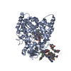
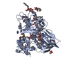
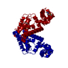
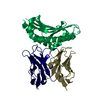
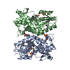
| Entry | Database: PDB / ID: 7dym | ||||||
|---|---|---|---|---|---|---|---|
| Title | Pseudomonas aeruginosa TseT-TsiT complex | ||||||
 Components Components |
| ||||||
 Keywords Keywords | TOXIN / T6SS / immunity / effector / nuclease | ||||||
| Function / homology | Tox-REase-5 domain / Restriction endonuclease fold toxin 5 / Imm52 family, TsiT-like / Immunity protein 52 / Immunity protein 52 / Immunity protein 52 domain-containing protein / Tox-REase-5 domain-containing protein Function and homology information Function and homology information | ||||||
| Biological species |   Pseudomonas aeruginosa (bacteria) Pseudomonas aeruginosa (bacteria) | ||||||
| Method |  X-RAY DIFFRACTION / SYNCHROTRON / SAD / Resolution: 3.1 Å X-RAY DIFFRACTION / SYNCHROTRON / SAD / Resolution: 3.1 Å | ||||||
 Authors Authors | She, Z. | ||||||
| Funding support |  China, 1items China, 1items
| ||||||
 Citation Citation | Journal: Int.J.Biol.Macromol. / Year: 2021 Title: Structure and SAXS studies unveiled a novel inhibition mechanism of the Pseudomonas aeruginosa T6SS TseT-TsiT complex. Authors: Wen, H. / Liu, G. / Geng, Z. / Zhang, H. / Li, Y. / She, Z. / Dong, Y. | ||||||
| History |
|
- Structure visualization
| Structure viewer | Molecule: MolmilJmol/JSmol |
|---|
- Downloads & links
-Download
| PDBx/mmCIF format | 7dym.cif.gz | 184.3 KB | Display | PDBx/mmCIF format |
|---|---|---|---|---|
| PDB format | pdb7dym.ent.gz | 147.1 KB | Display | PDB format |
| PDBx/mmJSON format | 7dym.json.gz | Tree view | PDBx/mmJSON format | |
| Others |  Other downloads Other downloads |
-Validation report
| Summary document | 7dym_validation.pdf.gz | 444.3 KB | Display | wwPDB validaton report |
|---|---|---|---|---|
| Full document | 7dym_full_validation.pdf.gz | 454.3 KB | Display | |
| Data in XML | 7dym_validation.xml.gz | 17.2 KB | Display | |
| Data in CIF | 7dym_validation.cif.gz | 22.2 KB | Display | |
| Arichive directory | https://data.pdbj.org/pub/pdb/validation_reports/dy/7dymftp://data.pdbj.org/pub/pdb/validation_reports/dy/7dym | HTTPS FTP |
-Related structure data
| Similar structure data |
|---|


-Links
 PDBj
PDBj- Assembly
| Deposited unit | 
| ||||||||
|---|---|---|---|---|---|---|---|---|---|
| 1 |
| ||||||||
| Unit cell |
|
-Components
| #1: Protein | Mass: 27708.621 Da / Num. of mol.: 1 Source method: isolated from a genetically manipulated source Source: (gene. exp.) Pseudomonas aeruginosa (strain ATCC 15692 / DSM 22644 / CIP 104116 / JCM 14847 / LMG 12228 / 1C / PRS 101 / PAO1) (bacteria)Strain: ATCC 15692 / DSM 22644 / CIP 104116 / JCM 14847 / LMG 12228 / 1C / PRS 101 / PAO1 Gene: PA3907 / Production host: |
|---|---|
| #2: Protein | Mass: 27328.992 Da / Num. of mol.: 1 Source method: isolated from a genetically manipulated source Source: (gene. exp.) Pseudomonas aeruginosa (strain ATCC 15692 / DSM 22644 / CIP 104116 / JCM 14847 / LMG 12228 / 1C / PRS 101 / PAO1) (bacteria)Strain: ATCC 15692 / DSM 22644 / CIP 104116 / JCM 14847 / LMG 12228 / 1C / PRS 101 / PAO1 Gene: PA3908 / Production host: |
| Has protein modification | Y |
-Experimental details
-Experiment
| Experiment | Method: X-RAY DIFFRACTION / Number of used crystals: 1 |
|---|
- Sample preparation
| Crystal | Density Matthews: 2.82 Å3/Da / Density % sol: 56.31 % |
|---|---|
| Crystal grow | Temperature: 293 K / Method: vapor diffusion, sitting drop / pH: 8.5 Details: 0.18 m Trimethylamine N-oxide, 0.1M Tris pH 8.5, 11% PEG2000MME |
-Data collection
| Diffraction | Mean temperature: 100 K / Serial crystal experiment: N |
|---|---|
| Diffraction source | Source: SYNCHROTRON / Site: SSRF / Beamline: BL17U1 / Wavelength: 0.9792 Å |
| Detector | Type: DECTRIS EIGER X 16M / Detector: PIXEL / Date: Jul 13, 2020 |
| Radiation | Protocol: SINGLE WAVELENGTH / Monochromatic (M) / Laue (L): M / Scattering type: x-ray |
| Radiation wavelength | Wavelength: 0.9792 Å / Relative weight: 1 |
| Reflection | Resolution: 3.1→45.53 Å / Num. obs: 11293 / % possible obs: 98.78 % / Redundancy: 10 % / CC1/2: 0.99 / Rmerge(I) obs: 0.063 / Net I/σ(I): 23.34 |
| Reflection shell | Resolution: 3.1→3.18 Å / Rmerge(I) obs: 0.48 / Num. unique obs: 777 / CC1/2: 0.96 |
- Processing
| Software |
| |||||||||||||||||||||||||||||||||||
|---|---|---|---|---|---|---|---|---|---|---|---|---|---|---|---|---|---|---|---|---|---|---|---|---|---|---|---|---|---|---|---|---|---|---|---|---|
| Refinement | Method to determine structure: SAD / Resolution: 3.1→25.37 Å / SU ML: 0.4 / Cross valid method: THROUGHOUT / σ(F): 1.42 / Phase error: 28.29 / Stereochemistry target values: ML
| |||||||||||||||||||||||||||||||||||
| Solvent computation | Shrinkage radii: 0.9 Å / VDW probe radii: 1.11 Å / Solvent model: FLAT BULK SOLVENT MODEL | |||||||||||||||||||||||||||||||||||
| Displacement parameters | Biso max: 220.63 Å2 / Biso mean: 111.2169 Å2 / Biso min: 56.97 Å2 | |||||||||||||||||||||||||||||||||||
| Refinement step | Cycle: final / Resolution: 3.1→25.37 Å
| |||||||||||||||||||||||||||||||||||
| LS refinement shell | Refine-ID: X-RAY DIFFRACTION / Rfactor Rfree error: 0 / Total num. of bins used: 4
|
