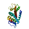
| Deposited unit | A: Endolysin
B: Endolysin
C: Endolysin
hetero molecules
| Theoretical mass | Number of molelcules |
|---|
| Total (without water) | 58,751 | 7 |
|---|
| Polymers | 58,489 | 3 |
|---|
| Non-polymers | 262 | 4 |
|---|
| Water | 162 | 9 |
|---|
|
|---|
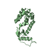
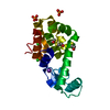
| 1 | A: Endolysin
hetero molecules
| Theoretical mass | Number of molelcules |
|---|
| Total (without water) | 19,562 | 2 |
|---|
| Polymers | 19,496 | 1 |
|---|
| Non-polymers | 65 | 1 |
|---|
| Water | 18 | 1 |
|---|
| Type | Name | Symmetry operation | Number |
|---|
| identity operation | 1_555 | x,y,z | 1 |
| Buried area | 0 Å2 |
|---|
| ΔGint | 0 kcal/mol |
|---|
| Surface area | 8510 Å2 |
|---|
| Method | PISA |
|---|
|
|---|
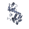
| 2 | B: Endolysin
hetero molecules
| Theoretical mass | Number of molelcules |
|---|
| Total (without water) | 19,627 | 3 |
|---|
| Polymers | 19,496 | 1 |
|---|
| Non-polymers | 131 | 2 |
|---|
| Water | 18 | 1 |
|---|
| Type | Name | Symmetry operation | Number |
|---|
| identity operation | 1_555 | x,y,z | 1 |
| Buried area | 60 Å2 |
|---|
| ΔGint | -19 kcal/mol |
|---|
| Surface area | 7720 Å2 |
|---|
| Method | PISA |
|---|
|
|---|
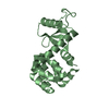
| 3 | C: Endolysin
hetero molecules
| Theoretical mass | Number of molelcules |
|---|
| Total (without water) | 19,562 | 2 |
|---|
| Polymers | 19,496 | 1 |
|---|
| Non-polymers | 65 | 1 |
|---|
| Water | 18 | 1 |
|---|
| Type | Name | Symmetry operation | Number |
|---|
| identity operation | 1_555 | x,y,z | 1 |
| Buried area | 60 Å2 |
|---|
| ΔGint | -17 kcal/mol |
|---|
| Surface area | 6620 Å2 |
|---|
| Method | PISA |
|---|
|
|---|
| Unit cell | | Length a, b, c (Å) | 102.861, 102.861, 244.533 |
|---|
| Angle α, β, γ (deg.) | 90.000, 90.000, 120.000 |
|---|
| Int Tables number | 180 |
|---|
| Space group name H-M | P6222 |
|---|
|
|---|
| Components on special symmetry positions | | ID | Model | Components |
|---|
| 1 | 1 | A-303- HOH | | 2 | 1 | A-305-HOH |
|
|---|
| Noncrystallographic symmetry (NCS) | NCS domain: | ID | Ens-ID | Details |
|---|
| 1 | 1 | (chain C and (resseq 0 or (resid 1 and (name...| 2 | 1 | (chain B and (resseq 0:12 or (resid 13 and (name... | |
NCS domain segments: Ens-ID: 1 | Dom-ID | Component-ID | Beg auth comp-ID | Beg label comp-ID | End auth comp-ID | End label comp-ID | Selection details | Auth asym-ID | Label asym-ID | Auth seq-ID | Label seq-ID |
|---|
| 1 | 1 | SERSERSERSER(chain C and (resseq 0 or (resid 1 and (name...CC| 0 | 5 | | 1 | 2 | METMETMETMET(chain C and (resseq 0 or (resid 1 and (name...CC| 1 | 6 | | 1 | 3 | SERSERLYSLYS(chain C and (resseq 0 or (resid 1 and (name...CC| 0 - 162 | 5 - 167 | | 1 | 4 | SERSERLYSLYS(chain C and (resseq 0 or (resid 1 and (name...CC| 0 - 162 | 5 - 167 | | 2 | 1 | SERSERGLYGLY(chain B and (resseq 0:12 or (resid 13 and (name...BB| 0 - 12 | 5 - 17 | | 2 | 2 | LEULEULEULEU(chain B and (resseq 0:12 or (resid 13 and (name...BB| 13 | 18 | | 2 | 3 | GLY | | | | | | | | | | | | | | | | | | | | | | | | | | | | | | | | | | | | | | | | | | |
|
|---|
 Movie
Movie Controller

 Yorodumi
 Open data
 Basic information
 Components
 Keywords
 Function and homology information
 Enterobacteria phage T4 (virus)
 X-RAY DIFFRACTION /
  Authors
 China, 1items
  Citation
 Structure visualization
 Downloads & links
 Other downloads
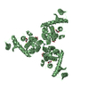


 PDBj
PDBj


 Assembly


