 Movie
Movie Controller


+ Open data

- Basic information
| Entry | Database: PDB / ID: 7dvj | ||||||
|---|---|---|---|---|---|---|---|
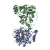
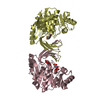
| Title | Structure of beta-mannanase BaMan113A with mannobiose | ||||||
 Components Components | Endo-beta-1,4-mannanase | ||||||
 Keywords Keywords | HYDROLASE / Complex | ||||||
| Function / homology | : / Glycoside Hydrolase Family 113 / Glycoside hydrolase superfamily / 4beta-beta-mannobiose / Endo-beta-1,4-mannanase Function and homology information Function and homology information | ||||||
| Biological species |  | ||||||
| Method |  X-RAY DIFFRACTION / SYNCHROTRON / MOLECULAR REPLACEMENT / Resolution: 1.65 Å X-RAY DIFFRACTION / SYNCHROTRON / MOLECULAR REPLACEMENT / Resolution: 1.65 Å | ||||||
 Authors Authors | Liu, W.T. / Liu, W.D. / Zheng, Y.Y. | ||||||
 Citation Citation | Journal: Int.J.Biol.Macromol. / Year: 2021 Title: Functional and structural investigation of a novel beta-mannanase BaMan113A from Bacillus sp. N16-5. Authors: Liu, W. / Ma, C. / Liu, W. / Zheng, Y. / Chen, C.C. / Liang, A. / Luo, X. / Li, Z. / Ma, W. / Song, Y. / Guo, R.T. / Zhang, T. | ||||||
| History |
|
- Structure visualization
| Structure viewer | Molecule: MolmilJmol/JSmol |
|---|
- Downloads & links
-Download
| PDBx/mmCIF format | 7dvj.cif.gz | 162.6 KB | Display | PDBx/mmCIF format |
|---|---|---|---|---|
| PDB format | pdb7dvj.ent.gz | 124.2 KB | Display | PDB format |
| PDBx/mmJSON format | 7dvj.json.gz | Tree view | PDBx/mmJSON format | |
| Others |  Other downloads Other downloads |
-Validation report
| Arichive directory | https://data.pdbj.org/pub/pdb/validation_reports/dv/7dvjftp://data.pdbj.org/pub/pdb/validation_reports/dv/7dvj | HTTPS FTP |
|---|
-Related structure data
| Related structure data |  7dv7C  7dvzC  7dw8C  7dwaC  3civS S: Starting model for refinement C: citing same article ( |
|---|---|
| Similar structure data |


-Links
 PDBj
PDBj- Assembly
| Deposited unit | 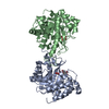
| ||||||||
|---|---|---|---|---|---|---|---|---|---|
| 1 |
| ||||||||
| Unit cell |
|
-Components
| #1: Protein | Mass: 40589.691 Da / Num. of mol.: 2 / Mutation: E142A Source method: isolated from a genetically manipulated source Source: (gene. exp.) #2: Polysaccharide |   Source method: isolated from a genetically manipulated source Details: oligosaccharide / References: 4beta-beta-mannobiose #3: Water | ChemComp-HOH / |  Mass: 18.015 Da / Num. of mol.: 764 / Source method: isolated from a natural source / Formula: H2O Mass: 18.015 Da / Num. of mol.: 764 / Source method: isolated from a natural source / Formula: H2OHas ligand of interest | Y | |
|---|
-Experimental details
-Experiment
| Experiment | Method: X-RAY DIFFRACTION / Number of used crystals: 1 |
|---|
- Sample preparation
| Crystal | Density Matthews: 2.89 Å3/Da / Density % sol: 57.39 % |
|---|---|
| Crystal grow | Temperature: 295 K / Method: vapor diffusion, sitting drop / Details: tacsimate pH 7.0 |
-Data collection
| Diffraction | Mean temperature: 100 K / Serial crystal experiment: N |
|---|---|
| Diffraction source | Source: SYNCHROTRON / Site: SSRF  / Beamline: BL19U1 / Wavelength: 0.979 Å / Beamline: BL19U1 / Wavelength: 0.979 Å |
| Detector | Type: DECTRIS PILATUS3 S 6M / Detector: PIXEL / Date: Oct 19, 2018 |
| Radiation | Protocol: SINGLE WAVELENGTH / Monochromatic (M) / Laue (L): M / Scattering type: x-ray |
| Radiation wavelength | Wavelength: 0.979 Å / Relative weight: 1 |
| Reflection | Resolution: 1.65→25 Å / Num. obs: 108815 / % possible obs: 97.9 % / Redundancy: 7.4 % / CC1/2: 0.858 / Net I/σ(I): 11.8 |
| Reflection shell | Resolution: 1.65→1.71 Å / Num. unique obs: 10962 / CC1/2: 0.905 |
- Processing
| Software |
| ||||||||||||||||||||||||||||||||||||||||||||||||||||||||||||
|---|---|---|---|---|---|---|---|---|---|---|---|---|---|---|---|---|---|---|---|---|---|---|---|---|---|---|---|---|---|---|---|---|---|---|---|---|---|---|---|---|---|---|---|---|---|---|---|---|---|---|---|---|---|---|---|---|---|---|---|---|---|
| Refinement | Method to determine structure: MOLECULAR REPLACEMENT Starting model: 3CIV Resolution: 1.65→25 Å / Cor.coef. Fo:Fc: 0.976 / Cor.coef. Fo:Fc free: 0.97 / SU B: 1.827 / SU ML: 0.058 / Cross valid method: THROUGHOUT / σ(F): 0 / ESU R: 0.078 / ESU R Free: 0.078 / Stereochemistry target values: MAXIMUM LIKELIHOOD Details: HYDROGENS HAVE BEEN ADDED IN THE RIDING POSITIONS U VALUES : REFINED INDIVIDUALLY
| ||||||||||||||||||||||||||||||||||||||||||||||||||||||||||||
| Solvent computation | Ion probe radii: 0.8 Å / Shrinkage radii: 0.8 Å / VDW probe radii: 1.2 Å / Solvent model: MASK | ||||||||||||||||||||||||||||||||||||||||||||||||||||||||||||
| Displacement parameters | Biso max: 111.35 Å2 / Biso mean: 21.568 Å2 / Biso min: 11.48 Å2
| ||||||||||||||||||||||||||||||||||||||||||||||||||||||||||||
| Refinement step | Cycle: final / Resolution: 1.65→25 Å
| ||||||||||||||||||||||||||||||||||||||||||||||||||||||||||||
| Refine LS restraints |
| ||||||||||||||||||||||||||||||||||||||||||||||||||||||||||||
| LS refinement shell | Resolution: 1.654→1.697 Å / Rfactor Rfree error: 0 / Total num. of bins used: 20
|
