 Movie
Movie Controller

[English] 日本語
 Yorodumi
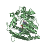
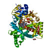
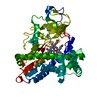
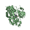
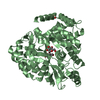
- PDB-7dtp: Crystal structure of agmatine coumaroyltransferase from Triticum ... -
+ Open data

- Basic information
| Entry | Database: PDB / ID: 7dtp | ||||||
|---|---|---|---|---|---|---|---|
| Title | Crystal structure of agmatine coumaroyltransferase from Triticum aestivum | ||||||
 Components Components | agmatine coumaroyltransferase | ||||||
 Keywords Keywords | TRANSFERASE / N-acyltransferase | ||||||
| Function / homology | : / Transferase family / acyltransferase activity, transferring groups other than amino-acyl groups / Chloramphenicol acetyltransferase-like domain superfamily / Agmatine coumaroyltransferase-2 Function and homology information Function and homology information | ||||||
| Biological species |  | ||||||
| Method |  X-RAY DIFFRACTION / SYNCHROTRON / MOLECULAR REPLACEMENT / Resolution: 2.3 Å X-RAY DIFFRACTION / SYNCHROTRON / MOLECULAR REPLACEMENT / Resolution: 2.3 Å | ||||||
 Authors Authors | Yamane, M. / Takenoya, M. / Sue, M. / Yajima, S. | ||||||
 Citation Citation | Journal: Phytochemistry / Year: 2021 Title: Molecular and structural characterization of agmatine coumaroyltransferase in Triticeae, the key regulator of hydroxycinnamic acid amide accumulation. Authors: Yamane, M. / Takenoya, M. / Yajima, S. / Sue, M. | ||||||
| History |
|
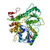
- Structure visualization
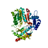
| Structure viewer | Molecule: MolmilJmol/JSmol |
|---|
- Downloads & links
-Download
| PDBx/mmCIF format | 7dtp.cif.gz | 98.8 KB | Display | PDBx/mmCIF format |
|---|---|---|---|---|
| PDB format | pdb7dtp.ent.gz | 69.7 KB | Display | PDB format |
| PDBx/mmJSON format | 7dtp.json.gz | Tree view | PDBx/mmJSON format | |
| Others |  Other downloads Other downloads |
-Validation report
| Summary document | 7dtp_validation.pdf.gz | 433.2 KB | Display | wwPDB validaton report |
|---|---|---|---|---|
| Full document | 7dtp_full_validation.pdf.gz | 435.3 KB | Display | |
| Data in XML | 7dtp_validation.xml.gz | 16.3 KB | Display | |
| Data in CIF | 7dtp_validation.cif.gz | 22.9 KB | Display | |
| Arichive directory | https://data.pdbj.org/pub/pdb/validation_reports/dt/7dtpftp://data.pdbj.org/pub/pdb/validation_reports/dt/7dtp | HTTPS FTP |
-Related structure data
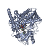
| Related structure data | 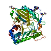 7cysS S: Starting model for refinement |
|---|---|
| Similar structure data |
-Links
 PDBj
PDBj

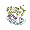
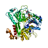
- Assembly
| Deposited unit | 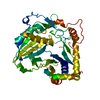
| ||||||||
|---|---|---|---|---|---|---|---|---|---|
| 1 |
| ||||||||
| Unit cell |
|
-Components
| #1: Protein | Mass: 51876.641 Da / Num. of mol.: 1 Source method: isolated from a genetically manipulated source Source: (gene. exp.)  |
|---|---|
| #2: Water | ChemComp-HOH /  Mass: 18.015 Da / Num. of mol.: 104 / Source method: isolated from a natural source / Formula: H2O Mass: 18.015 Da / Num. of mol.: 104 / Source method: isolated from a natural source / Formula: H2O |
| Has protein modification | Y |
-Experimental details
-Experiment
| Experiment | Method: X-RAY DIFFRACTION / Number of used crystals: 1 |
|---|
- Sample preparation
| Crystal | Density Matthews: 2.17 Å3/Da / Density % sol: 43.26 % |
|---|---|
| Crystal grow | Temperature: 293 K / Method: vapor diffusion, hanging drop / pH: 6.5 / Details: 100 mM MMT buffer pH 6.5, 25 % (w/v) PEG 1500 |
-Data collection
| Diffraction | Mean temperature: 95 K / Serial crystal experiment: N |
|---|---|
| Diffraction source | Source: SYNCHROTRON / Site: Photon Factory  / Beamline: AR-NW12A / Wavelength: 1 Å / Beamline: AR-NW12A / Wavelength: 1 Å |
| Detector | Type: DECTRIS PILATUS3 S 6M / Detector: PIXEL / Date: Oct 30, 2020 |
| Radiation | Protocol: SINGLE WAVELENGTH / Monochromatic (M) / Laue (L): M / Scattering type: x-ray |
| Radiation wavelength | Wavelength: 1 Å / Relative weight: 1 |
| Reflection | Resolution: 2.3→50 Å / Num. obs: 20776 / % possible obs: 100 % / Redundancy: 6.4 % / CC1/2: 0.993 / Rmerge(I) obs: 0.131 / Net I/σ(I): 14.2 |
| Reflection shell | Resolution: 2.3→2.34 Å / Redundancy: 6.6 % / Rmerge(I) obs: 0.927 / Mean I/σ(I) obs: 1.9 / Num. unique obs: 1033 / CC1/2: 0.782 / % possible all: 100 |
- Processing
| Software |
| |||||||||||||||||||||||||||||||||||||||||||||||||||||||||||||||||||||||||||||||||||||||||||||||||||||||||||||||||||||||||||||||||||||||||||||||||||||||||||
|---|---|---|---|---|---|---|---|---|---|---|---|---|---|---|---|---|---|---|---|---|---|---|---|---|---|---|---|---|---|---|---|---|---|---|---|---|---|---|---|---|---|---|---|---|---|---|---|---|---|---|---|---|---|---|---|---|---|---|---|---|---|---|---|---|---|---|---|---|---|---|---|---|---|---|---|---|---|---|---|---|---|---|---|---|---|---|---|---|---|---|---|---|---|---|---|---|---|---|---|---|---|---|---|---|---|---|---|---|---|---|---|---|---|---|---|---|---|---|---|---|---|---|---|---|---|---|---|---|---|---|---|---|---|---|---|---|---|---|---|---|---|---|---|---|---|---|---|---|---|---|---|---|---|---|---|---|
| Refinement | Method to determine structure: MOLECULAR REPLACEMENT Starting model: 7CYS Resolution: 2.3→48.948 Å / Cor.coef. Fo:Fc: 0.925 / Cor.coef. Fo:Fc free: 0.912 / SU B: 8.747 / SU ML: 0.208 / Cross valid method: THROUGHOUT / ESU R: 0.377 / ESU R Free: 0.243 Details: Hydrogens have been added in their riding positions
| |||||||||||||||||||||||||||||||||||||||||||||||||||||||||||||||||||||||||||||||||||||||||||||||||||||||||||||||||||||||||||||||||||||||||||||||||||||||||||
| Solvent computation | Ion probe radii: 0.8 Å / Shrinkage radii: 0.8 Å / VDW probe radii: 1.2 Å / Solvent model: MASK BULK SOLVENT | |||||||||||||||||||||||||||||||||||||||||||||||||||||||||||||||||||||||||||||||||||||||||||||||||||||||||||||||||||||||||||||||||||||||||||||||||||||||||||
| Displacement parameters | Biso mean: 37.97 Å2
| |||||||||||||||||||||||||||||||||||||||||||||||||||||||||||||||||||||||||||||||||||||||||||||||||||||||||||||||||||||||||||||||||||||||||||||||||||||||||||
| Refinement step | Cycle: LAST / Resolution: 2.3→48.948 Å
| |||||||||||||||||||||||||||||||||||||||||||||||||||||||||||||||||||||||||||||||||||||||||||||||||||||||||||||||||||||||||||||||||||||||||||||||||||||||||||
| Refine LS restraints |
| |||||||||||||||||||||||||||||||||||||||||||||||||||||||||||||||||||||||||||||||||||||||||||||||||||||||||||||||||||||||||||||||||||||||||||||||||||||||||||
| LS refinement shell |
|
