 Movie
Movie Controller

[English] 日本語
 Yorodumi
- PDB-7cys: Crystal structure of barley agmatine coumaroyltransferase (HvACT)... -
+ Open data

- Basic information
| Entry | Database: PDB / ID: 7cys | ||||||
|---|---|---|---|---|---|---|---|
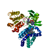
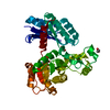
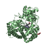
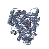
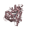
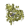
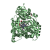
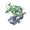
| Title | Crystal structure of barley agmatine coumaroyltransferase (HvACT), an N-acyltransferase in BAHD superfamily | ||||||
 Components Components | Agmatine coumaroyltransferase-1 | ||||||
 Keywords Keywords | TRANSFERASE / acyltransferase | ||||||
| Function / homology | agmatine N4-coumaroyltransferase / agmatine N4-coumaroyltransferase activity / : / Transferase family / Chloramphenicol acetyltransferase-like domain superfamily / Agmatine coumaroyltransferase-1 Function and homology information Function and homology information | ||||||
| Biological species |  | ||||||
| Method |  X-RAY DIFFRACTION / SYNCHROTRON / MOLECULAR REPLACEMENT / Resolution: 1.81 Å X-RAY DIFFRACTION / SYNCHROTRON / MOLECULAR REPLACEMENT / Resolution: 1.81 Å | ||||||
 Authors Authors | Yamane, M. / Takenoya, M. / Sue, M. / Yajima, S. | ||||||
 Citation Citation | Journal: Acta Crystallogr.,Sect.F / Year: 2020 Title: Crystal structure of barley agmatine coumaroyltransferase, an N-acyltransferase from the BAHD superfamily. Authors: Yamane, M. / Takenoya, M. / Yajima, S. / Sue, M. | ||||||
| History |
|
- Structure visualization
| Structure viewer | Molecule: MolmilJmol/JSmol |
|---|
- Downloads & links
-Download
| PDBx/mmCIF format | 7cys.cif.gz | 106.7 KB | Display | PDBx/mmCIF format |
|---|---|---|---|---|
| PDB format | pdb7cys.ent.gz | 76 KB | Display | PDB format |
| PDBx/mmJSON format | 7cys.json.gz | Tree view | PDBx/mmJSON format | |
| Others |  Other downloads Other downloads |
-Validation report
| Arichive directory | https://data.pdbj.org/pub/pdb/validation_reports/cy/7cysftp://data.pdbj.org/pub/pdb/validation_reports/cy/7cys | HTTPS FTP |
|---|
-Related structure data
| Related structure data | 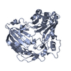 6dd2S S: Starting model for refinement |
|---|---|
| Similar structure data |


-Links
 PDBj
PDBj

- Assembly
| Deposited unit | 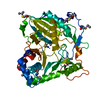
| ||||||||
|---|---|---|---|---|---|---|---|---|---|
| 1 |
| ||||||||
| Unit cell |
|
-Components
| #1: Protein | Mass: 51628.312 Da / Num. of mol.: 1 Source method: isolated from a genetically manipulated source Source: (gene. exp.)  References: UniProt: A9ZPJ6, agmatine N4-coumaroyltransferase |
|---|---|
| #2: Water | ChemComp-HOH /  Mass: 18.015 Da / Num. of mol.: 226 / Source method: isolated from a natural source / Formula: H2O Mass: 18.015 Da / Num. of mol.: 226 / Source method: isolated from a natural source / Formula: H2O |
| Has ligand of interest | N |
| Has protein modification | Y |
-Experimental details
-Experiment
| Experiment | Method: X-RAY DIFFRACTION / Number of used crystals: 1 |
|---|
- Sample preparation
| Crystal | Density Matthews: 2.45 Å3/Da / Density % sol: 49.7 % |
|---|---|
| Crystal grow | Temperature: 293 K / Method: vapor diffusion, hanging drop / pH: 6.5 Details: 0.08 M sodium cacodylate, pH 6.5, 0.16 M magnesium acetate, 16% (w/v) polyethylene glycol 8,000, and 20% (v/v) glycerol |
-Data collection
| Diffraction | Mean temperature: 95 K / Serial crystal experiment: N |
|---|---|
| Diffraction source | Source: SYNCHROTRON / Site: Photon Factory  / Beamline: BL-5A / Wavelength: 1 Å / Beamline: BL-5A / Wavelength: 1 Å |
| Detector | Type: DECTRIS PILATUS3 S 6M / Detector: PIXEL / Date: Jun 23, 2020 |
| Radiation | Protocol: SINGLE WAVELENGTH / Monochromatic (M) / Laue (L): M / Scattering type: x-ray |
| Radiation wavelength | Wavelength: 1 Å / Relative weight: 1 |
| Reflection | Resolution: 1.81→50 Å / Num. obs: 44594 / % possible obs: 97.8 % / Redundancy: 3.1 % / CC1/2: 0.999 / Rmerge(I) obs: 0.036 / Net I/σ(I): 16.3 |
| Reflection shell | Resolution: 1.81→1.85 Å / Redundancy: 2 % / Mean I/σ(I) obs: 2.2 / Num. unique obs: 2201 / CC1/2: 0.914 / CC star: 0.237 / % possible all: 82.5 |
- Processing
| Software |
| ||||||||||||||||||||||||||||||||||||||||||||||||||||||||||||||||||||||||||||||||||||||||||||||||||||||||||||||||||||||||||||||||||||||||||||||||||||||
|---|---|---|---|---|---|---|---|---|---|---|---|---|---|---|---|---|---|---|---|---|---|---|---|---|---|---|---|---|---|---|---|---|---|---|---|---|---|---|---|---|---|---|---|---|---|---|---|---|---|---|---|---|---|---|---|---|---|---|---|---|---|---|---|---|---|---|---|---|---|---|---|---|---|---|---|---|---|---|---|---|---|---|---|---|---|---|---|---|---|---|---|---|---|---|---|---|---|---|---|---|---|---|---|---|---|---|---|---|---|---|---|---|---|---|---|---|---|---|---|---|---|---|---|---|---|---|---|---|---|---|---|---|---|---|---|---|---|---|---|---|---|---|---|---|---|---|---|---|---|---|---|
| Refinement | Method to determine structure: MOLECULAR REPLACEMENT Starting model: 6dd2 Resolution: 1.81→46.326 Å / Cor.coef. Fo:Fc: 0.95 / Cor.coef. Fo:Fc free: 0.951 / SU B: 2.544 / SU ML: 0.077 / Cross valid method: THROUGHOUT / ESU R: 0.125 / ESU R Free: 0.109 Details: Hydrogens have been added in their riding positions
| ||||||||||||||||||||||||||||||||||||||||||||||||||||||||||||||||||||||||||||||||||||||||||||||||||||||||||||||||||||||||||||||||||||||||||||||||||||||
| Solvent computation | Ion probe radii: 0.8 Å / Shrinkage radii: 0.8 Å / VDW probe radii: 1.2 Å / Solvent model: MASK BULK SOLVENT | ||||||||||||||||||||||||||||||||||||||||||||||||||||||||||||||||||||||||||||||||||||||||||||||||||||||||||||||||||||||||||||||||||||||||||||||||||||||
| Displacement parameters | Biso mean: 23.607 Å2
| ||||||||||||||||||||||||||||||||||||||||||||||||||||||||||||||||||||||||||||||||||||||||||||||||||||||||||||||||||||||||||||||||||||||||||||||||||||||
| Refinement step | Cycle: LAST / Resolution: 1.81→46.326 Å
| ||||||||||||||||||||||||||||||||||||||||||||||||||||||||||||||||||||||||||||||||||||||||||||||||||||||||||||||||||||||||||||||||||||||||||||||||||||||
| Refine LS restraints |
| ||||||||||||||||||||||||||||||||||||||||||||||||||||||||||||||||||||||||||||||||||||||||||||||||||||||||||||||||||||||||||||||||||||||||||||||||||||||
| LS refinement shell |
|
