[English] 日本語
 Yorodumi
- PDB-7ds2: Crystal structure of actin capping protein in complex with twinfl... -
+ Open data
- Basic information
| Entry | Database: PDB / ID: 7ds2 | ||||||||||||
|---|---|---|---|---|---|---|---|---|---|---|---|---|---|
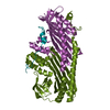
| Title | Crystal structure of actin capping protein in complex with twinflin-1 C-terminus tail | ||||||||||||
 Components Components |
| ||||||||||||
 Keywords Keywords | CYTOSOLIC PROTEIN / actin dynamics / actin capping protein / twinfilin / CARMIL / V-1 | ||||||||||||
| Function / homology |  Function and homology information Function and homology informationAdvanced glycosylation endproduct receptor signaling / RHOD GTPase cycle / RHOF GTPase cycle / HSP90 chaperone cycle for steroid hormone receptors (SHR) in the presence of ligand / COPI-independent Golgi-to-ER retrograde traffic / RHOBTB2 GTPase cycle / Factors involved in megakaryocyte development and platelet production / COPI-mediated anterograde transport / negative regulation of filopodium assembly / sperm head-tail coupling apparatus ...Advanced glycosylation endproduct receptor signaling / RHOD GTPase cycle / RHOF GTPase cycle / HSP90 chaperone cycle for steroid hormone receptors (SHR) in the presence of ligand / COPI-independent Golgi-to-ER retrograde traffic / RHOBTB2 GTPase cycle / Factors involved in megakaryocyte development and platelet production / COPI-mediated anterograde transport / negative regulation of filopodium assembly / sperm head-tail coupling apparatus / F-actin capping protein complex / WASH complex / negative regulation of actin filament polymerization / cell junction assembly / barbed-end actin filament capping / actin polymerization or depolymerization / regulation of lamellipodium assembly / regulation of cell morphogenesis / lamellipodium assembly / positive regulation of cardiac muscle hypertrophy / cortical cytoskeleton / myofibril / brush border / actin monomer binding / phosphatidylinositol-4,5-bisphosphate binding / cytoskeleton organization / hippocampal mossy fiber to CA3 synapse / filopodium / Schaffer collateral - CA1 synapse / Z disc / cell morphogenesis / actin filament binding / cell-cell junction / lamellipodium / actin cytoskeleton / actin cytoskeleton organization / protein tyrosine kinase activity / postsynaptic density / protein-containing complex binding / perinuclear region of cytoplasm / ATP binding / membrane / plasma membrane / cytosol Similarity search - Function | ||||||||||||
| Biological species |   | ||||||||||||
| Method |  X-RAY DIFFRACTION / X-RAY DIFFRACTION /  MOLECULAR REPLACEMENT / Resolution: 1.95 Å MOLECULAR REPLACEMENT / Resolution: 1.95 Å | ||||||||||||
 Authors Authors | Takeda, S. | ||||||||||||
| Funding support |  Japan, 3items Japan, 3items
| ||||||||||||
 Citation Citation |  Journal: J.Mol.Biol. / Year: 2021 Journal: J.Mol.Biol. / Year: 2021Title: Structural Insights into the Regulation of Actin Capping Protein by Twinfilin C-terminal Tail. Authors: Takeda, S. / Koike, R. / Fujiwara, I. / Narita, A. / Miyata, M. / Ota, M. / Maeda, Y. | ||||||||||||
| History |
|
- Structure visualization
| Structure viewer | Molecule:  Molmil Molmil Jmol/JSmol Jmol/JSmol |
|---|
- Downloads & links
- Download
| PDBx/mmCIF format |  7ds2.cif.gz 7ds2.cif.gz | 146.7 KB | Display |  PDBx/mmCIF format PDBx/mmCIF format |
|---|---|---|---|---|
| PDB format |  pdb7ds2.ent.gz pdb7ds2.ent.gz | 102.3 KB | Display |  PDB format PDB format |
| PDBx/mmJSON format |  7ds2.json.gz 7ds2.json.gz | Tree view |  PDBx/mmJSON format PDBx/mmJSON format | |
| Others |  Other downloads Other downloads |
-Validation report
| Summary document |  7ds2_validation.pdf.gz 7ds2_validation.pdf.gz | 438.2 KB | Display |  wwPDB validaton report wwPDB validaton report |
|---|---|---|---|---|
| Full document |  7ds2_full_validation.pdf.gz 7ds2_full_validation.pdf.gz | 440 KB | Display | |
| Data in XML |  7ds2_validation.xml.gz 7ds2_validation.xml.gz | 25.4 KB | Display | |
| Data in CIF |  7ds2_validation.cif.gz 7ds2_validation.cif.gz | 37.4 KB | Display | |
| Arichive directory |  https://data.pdbj.org/pub/pdb/validation_reports/ds/7ds2 https://data.pdbj.org/pub/pdb/validation_reports/ds/7ds2 ftp://data.pdbj.org/pub/pdb/validation_reports/ds/7ds2 ftp://data.pdbj.org/pub/pdb/validation_reports/ds/7ds2 | HTTPS FTP |
-Related structure data
| Related structure data | 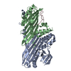 7ds3C 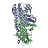 7ds4C 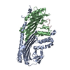 7ds6C 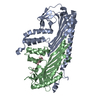 7ds8C 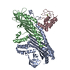 7dsaC 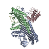 7dsbC 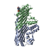 3aa7S S: Starting model for refinement C: citing same article ( |
|---|---|
| Similar structure data |
- Links
Links
- Assembly
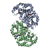
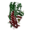
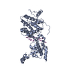
| Deposited unit | 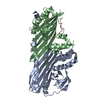
| ||||||||||||
|---|---|---|---|---|---|---|---|---|---|---|---|---|---|
| 1 |
| ||||||||||||
| Unit cell |
|
- Components
| #1: Protein | Mass: 33001.789 Da / Num. of mol.: 1 Source method: isolated from a genetically manipulated source Source: (gene. exp.)   |
|---|---|
| #2: Protein | Mass: 27473.070 Da / Num. of mol.: 1 Source method: isolated from a genetically manipulated source Source: (gene. exp.)   |
| #3: Protein/peptide | Mass: 4331.960 Da / Num. of mol.: 1 Source method: isolated from a genetically manipulated source Source: (gene. exp.)   |
| #4: Water | ChemComp-HOH / |
-Experimental details
-Experiment
| Experiment | Method:  X-RAY DIFFRACTION / Number of used crystals: 1 X-RAY DIFFRACTION / Number of used crystals: 1 |
|---|
- Sample preparation
| Crystal | Density Matthews: 1.99 Å3/Da / Density % sol: 38.34 % |
|---|---|
| Crystal grow | Temperature: 293 K / Method: vapor diffusion, hanging drop / pH: 7 / Details: 22.5% (w/v) PEG 400, 0.1M HEPES-NaOH (pH = 7.0) |
-Data collection
| Diffraction | Mean temperature: 95 K / Serial crystal experiment: N | ||||||||||||||||||||||||||||||||||||||||||||||||||||||||||||||||||||||||||||||||
|---|---|---|---|---|---|---|---|---|---|---|---|---|---|---|---|---|---|---|---|---|---|---|---|---|---|---|---|---|---|---|---|---|---|---|---|---|---|---|---|---|---|---|---|---|---|---|---|---|---|---|---|---|---|---|---|---|---|---|---|---|---|---|---|---|---|---|---|---|---|---|---|---|---|---|---|---|---|---|---|---|---|
| Diffraction source | Source:  ROTATING ANODE / Type: RIGAKU FR-E SUPERBRIGHT / Wavelength: 1.5418 Å ROTATING ANODE / Type: RIGAKU FR-E SUPERBRIGHT / Wavelength: 1.5418 Å | ||||||||||||||||||||||||||||||||||||||||||||||||||||||||||||||||||||||||||||||||
| Detector | Type: RIGAKU RAXIS IV / Detector: IMAGE PLATE / Date: Dec 26, 2012 | ||||||||||||||||||||||||||||||||||||||||||||||||||||||||||||||||||||||||||||||||
| Radiation | Protocol: SINGLE WAVELENGTH / Monochromatic (M) / Laue (L): M / Scattering type: x-ray | ||||||||||||||||||||||||||||||||||||||||||||||||||||||||||||||||||||||||||||||||
| Radiation wavelength | Wavelength: 1.5418 Å / Relative weight: 1 | ||||||||||||||||||||||||||||||||||||||||||||||||||||||||||||||||||||||||||||||||
| Reflection | Resolution: 1.95→48.97 Å / Num. obs: 37412 / % possible obs: 96.5 % / Redundancy: 7.109 % / Biso Wilson estimate: 31.802 Å2 / CC1/2: 1 / Rmerge(I) obs: 0.041 / Rrim(I) all: 0.044 / Χ2: 0.971 / Net I/σ(I): 31.64 | ||||||||||||||||||||||||||||||||||||||||||||||||||||||||||||||||||||||||||||||||
| Reflection shell | Diffraction-ID: 1
|
- Processing
| Software |
| ||||||||||||||||||||||||||||||||||||||||||||||||||||||||||||||||||||||||||||||||||||||||||||||||||
|---|---|---|---|---|---|---|---|---|---|---|---|---|---|---|---|---|---|---|---|---|---|---|---|---|---|---|---|---|---|---|---|---|---|---|---|---|---|---|---|---|---|---|---|---|---|---|---|---|---|---|---|---|---|---|---|---|---|---|---|---|---|---|---|---|---|---|---|---|---|---|---|---|---|---|---|---|---|---|---|---|---|---|---|---|---|---|---|---|---|---|---|---|---|---|---|---|---|---|---|
| Refinement | Method to determine structure:  MOLECULAR REPLACEMENT MOLECULAR REPLACEMENTStarting model: 3aa7 Resolution: 1.95→48.94 Å / SU ML: 0.1557 / Cross valid method: FREE R-VALUE / σ(F): 1.36 / Phase error: 17.2733 Stereochemistry target values: GeoStd + Monomer Library + CDL v1.2
| ||||||||||||||||||||||||||||||||||||||||||||||||||||||||||||||||||||||||||||||||||||||||||||||||||
| Solvent computation | Shrinkage radii: 0.9 Å / VDW probe radii: 1.11 Å / Solvent model: FLAT BULK SOLVENT MODEL | ||||||||||||||||||||||||||||||||||||||||||||||||||||||||||||||||||||||||||||||||||||||||||||||||||
| Displacement parameters | Biso mean: 28.43 Å2 | ||||||||||||||||||||||||||||||||||||||||||||||||||||||||||||||||||||||||||||||||||||||||||||||||||
| Refinement step | Cycle: LAST / Resolution: 1.95→48.94 Å
| ||||||||||||||||||||||||||||||||||||||||||||||||||||||||||||||||||||||||||||||||||||||||||||||||||
| Refine LS restraints |
| ||||||||||||||||||||||||||||||||||||||||||||||||||||||||||||||||||||||||||||||||||||||||||||||||||
| LS refinement shell |
|
 Movie
Movie Controller



 PDBj
PDBj

