 Movie
Movie Controller

[English] 日本語
 Yorodumi
- PDB-7drn: Structure of ATP-grasp ligase PsnB complexed with precursor pepti... -
+ Open data

- Basic information
| Entry | Database: PDB / ID: 7drn | ||||||
|---|---|---|---|---|---|---|---|
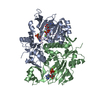
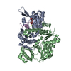
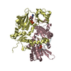
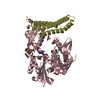
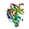
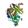
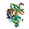
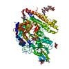
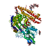
| Title | Structure of ATP-grasp ligase PsnB complexed with precursor peptide PsnA2 and AMPPNP | ||||||
 Components Components |
| ||||||
 Keywords Keywords | LIGASE / ATP-grasp ligase / RiPP / graspetide / Omega ester bond containing peptide | ||||||
| Function / homology |  Function and homology information Function and homology informationligase activity, forming carbon-nitrogen bonds / ATP binding / metal ion binding / cytoplasm Similarity search - Function | ||||||
| Biological species |  Plesiocystis pacifica SIR-1 (bacteria) Plesiocystis pacifica SIR-1 (bacteria) | ||||||
| Method |  X-RAY DIFFRACTION / SYNCHROTRON / MOLECULAR REPLACEMENT / Resolution: 3.56 Å X-RAY DIFFRACTION / SYNCHROTRON / MOLECULAR REPLACEMENT / Resolution: 3.56 Å | ||||||
 Authors Authors | Song, I. / Yu, J. / Song, W. / Kim, S. | ||||||
 Citation Citation | Journal: Nat.Chem.Biol. / Year: 2021 Title: Molecular mechanism underlying substrate recognition of the peptide macrocyclase PsnB. Authors: Song, I. / Kim, Y. / Yu, J. / Go, S.Y. / Lee, H.G. / Song, W.J. / Kim, S. | ||||||
| History |
|
- Structure visualization
| Structure viewer | Molecule: MolmilJmol/JSmol |
|---|
- Downloads & links
-Download
| PDBx/mmCIF format | 7drn.cif.gz | 478.8 KB | Display | PDBx/mmCIF format |
|---|---|---|---|---|
| PDB format | pdb7drn.ent.gz | 390.3 KB | Display | PDB format |
| PDBx/mmJSON format | 7drn.json.gz | Tree view | PDBx/mmJSON format | |
| Others |  Other downloads Other downloads |
-Validation report
| Arichive directory | https://data.pdbj.org/pub/pdb/validation_reports/dr/7drnftp://data.pdbj.org/pub/pdb/validation_reports/dr/7drn | HTTPS FTP |
|---|
-Related structure data
| Related structure data |  7drmC  7droC  7drpC  5ig9S S: Starting model for refinement C: citing same article ( |
|---|---|
| Similar structure data |
-Links
 PDBj
PDBj


- Assembly
| Deposited unit | 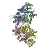
| ||||||||
|---|---|---|---|---|---|---|---|---|---|
| 1 | 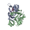
| ||||||||
| 2 | 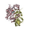
| ||||||||
| Unit cell |
|
-Components
| #1: Protein | Mass: 37023.816 Da / Num. of mol.: 4 Source method: isolated from a genetically manipulated source Source: (gene. exp.) Plesiocystis pacifica SIR-1 (bacteria) / Gene: PPSIR1_03893 / Production host: #2: Protein/peptide | Mass: 2567.886 Da / Num. of mol.: 2 / Source method: obtained synthetically / Source: (synth.) Plesiocystis pacifica SIR-1 (bacteria) / References: UniProt: A6GH40#3: Chemical |   Mass: 506.196 Da / Num. of mol.: 2 / Source method: obtained synthetically / Formula: C10H17N6O12P3 / Feature type: SUBJECT OF INVESTIGATION / Comment: AMP-PNP, energy-carrying molecule analogue*YM Mass: 506.196 Da / Num. of mol.: 2 / Source method: obtained synthetically / Formula: C10H17N6O12P3 / Feature type: SUBJECT OF INVESTIGATION / Comment: AMP-PNP, energy-carrying molecule analogue*YMHas ligand of interest | Y | |
|---|
-Experimental details
-Experiment
| Experiment | Method: X-RAY DIFFRACTION / Number of used crystals: 1 |
|---|
- Sample preparation
| Crystal | Density Matthews: 2.73 Å3/Da / Density % sol: 54.91 % |
|---|---|
| Crystal grow | Temperature: 293 K / Method: vapor diffusion, hanging drop / pH: 5.2 / Details: Sodium acetate, PEG3350 |
-Data collection
| Diffraction | Mean temperature: 100 K / Serial crystal experiment: N | |||||||||||||||||||||||||||||||||||||||||||||||||||||||||||||||||||||||||||||||||||||||||||||||||||||||||||||||||||||||||||||||||||||||||||||||||||||||||||||||||||||||||||||||||||||||||||||
|---|---|---|---|---|---|---|---|---|---|---|---|---|---|---|---|---|---|---|---|---|---|---|---|---|---|---|---|---|---|---|---|---|---|---|---|---|---|---|---|---|---|---|---|---|---|---|---|---|---|---|---|---|---|---|---|---|---|---|---|---|---|---|---|---|---|---|---|---|---|---|---|---|---|---|---|---|---|---|---|---|---|---|---|---|---|---|---|---|---|---|---|---|---|---|---|---|---|---|---|---|---|---|---|---|---|---|---|---|---|---|---|---|---|---|---|---|---|---|---|---|---|---|---|---|---|---|---|---|---|---|---|---|---|---|---|---|---|---|---|---|---|---|---|---|---|---|---|---|---|---|---|---|---|---|---|---|---|---|---|---|---|---|---|---|---|---|---|---|---|---|---|---|---|---|---|---|---|---|---|---|---|---|---|---|---|---|---|---|---|---|
| Diffraction source | Source: SYNCHROTRON / Site: PAL/PLS  / Beamline: 7A (6B, 6C1) / Wavelength: 0.97934 Å / Beamline: 7A (6B, 6C1) / Wavelength: 0.97934 Å | |||||||||||||||||||||||||||||||||||||||||||||||||||||||||||||||||||||||||||||||||||||||||||||||||||||||||||||||||||||||||||||||||||||||||||||||||||||||||||||||||||||||||||||||||||||||||||||
| Detector | Type: ADSC QUANTUM 270 / Detector: CCD / Date: Oct 18, 2018 | |||||||||||||||||||||||||||||||||||||||||||||||||||||||||||||||||||||||||||||||||||||||||||||||||||||||||||||||||||||||||||||||||||||||||||||||||||||||||||||||||||||||||||||||||||||||||||||
| Radiation | Protocol: SINGLE WAVELENGTH / Monochromatic (M) / Laue (L): M / Scattering type: x-ray | |||||||||||||||||||||||||||||||||||||||||||||||||||||||||||||||||||||||||||||||||||||||||||||||||||||||||||||||||||||||||||||||||||||||||||||||||||||||||||||||||||||||||||||||||||||||||||||
| Radiation wavelength | Wavelength: 0.97934 Å / Relative weight: 1 | |||||||||||||||||||||||||||||||||||||||||||||||||||||||||||||||||||||||||||||||||||||||||||||||||||||||||||||||||||||||||||||||||||||||||||||||||||||||||||||||||||||||||||||||||||||||||||||
| Reflection | Resolution: 3.56→50 Å / Num. obs: 19860 / % possible obs: 99.9 % / Redundancy: 7.4 % / Biso Wilson estimate: 72.41 Å2 / Rmerge(I) obs: 0.183 / Rpim(I) all: 0.072 / Rrim(I) all: 0.197 / Χ2: 1.942 / Net I/σ(I): 5.7 | |||||||||||||||||||||||||||||||||||||||||||||||||||||||||||||||||||||||||||||||||||||||||||||||||||||||||||||||||||||||||||||||||||||||||||||||||||||||||||||||||||||||||||||||||||||||||||||
| Reflection shell | Diffraction-ID: 1
|
- Processing
| Software |
| ||||||||||||||||||||||||||||||||||||||||||||||||||||||||
|---|---|---|---|---|---|---|---|---|---|---|---|---|---|---|---|---|---|---|---|---|---|---|---|---|---|---|---|---|---|---|---|---|---|---|---|---|---|---|---|---|---|---|---|---|---|---|---|---|---|---|---|---|---|---|---|---|---|
| Refinement | Method to determine structure: MOLECULAR REPLACEMENT Starting model: 5IG9 Resolution: 3.56→32.95 Å / SU ML: 0.34 / Cross valid method: THROUGHOUT / σ(F): 1.33 / Phase error: 24.13 / Stereochemistry target values: ML
| ||||||||||||||||||||||||||||||||||||||||||||||||||||||||
| Solvent computation | Shrinkage radii: 0.9 Å / VDW probe radii: 1.11 Å / Solvent model: FLAT BULK SOLVENT MODEL | ||||||||||||||||||||||||||||||||||||||||||||||||||||||||
| Displacement parameters | Biso max: 124.35 Å2 / Biso mean: 69.2455 Å2 / Biso min: 31.34 Å2 | ||||||||||||||||||||||||||||||||||||||||||||||||||||||||
| Refinement step | Cycle: final / Resolution: 3.56→32.95 Å
| ||||||||||||||||||||||||||||||||||||||||||||||||||||||||
| LS refinement shell | Refine-ID: X-RAY DIFFRACTION / Rfactor Rfree error: 0 / Total num. of bins used: 7
|
