 Movie
Movie Controller

[English] 日本語
 Yorodumi
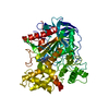
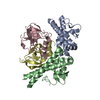
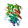
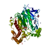
- PDB-7drk: Crystal structure of phosphatidylglycerol phosphate synthase in c... -
+ Open data

- Basic information
| Entry | Database: PDB / ID: 7drk | |||||||||
|---|---|---|---|---|---|---|---|---|---|---|
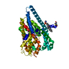
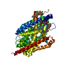
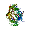
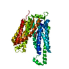
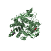
| Title | Crystal structure of phosphatidylglycerol phosphate synthase in complex with cytidine diphosphate-diacylglycerol | |||||||||
 Components Components | CDP-diacylglycerol--glycerol-3-phosphate 3-phosphatidyltransferase | |||||||||
 Keywords Keywords | TRANSFERASE / Substrate complex / Phospholipid synthase / Staphylococcus aureus / Transferase. | |||||||||
| Function / homology |  Function and homology information Function and homology informationCDP-diacylglycerol-glycerol-3-phosphate 1-phosphatidyltransferase / CDP-diacylglycerol-glycerol-3-phosphate 3-phosphatidyltransferase activity / phosphatidylglycerol biosynthetic process / plasma membrane Similarity search - Function | |||||||||
| Biological species |   Staphylococcus aureus (bacteria) Staphylococcus aureus (bacteria) | |||||||||
| Method |  X-RAY DIFFRACTION / SYNCHROTRON / MOLECULAR REPLACEMENT / Resolution: 3 Å X-RAY DIFFRACTION / SYNCHROTRON / MOLECULAR REPLACEMENT / Resolution: 3 Å | |||||||||
 Authors Authors | Yang, B.W. / Liu, Z.F. | |||||||||
| Funding support |  China, 2items China, 2items
| |||||||||
 Citation Citation | Journal: Curr Res Struct Biol / Year: 2021 Title: The phosphatidylglycerol phosphate synthase PgsA utilizes a trifurcated amphipathic cavity for catalysis at the membrane-cytosol interface. Authors: Yang, B. / Yao, H. / Li, D. / Liu, Z. | |||||||||
| History |
|
- Structure visualization
| Structure viewer | Molecule: MolmilJmol/JSmol |
|---|
- Downloads & links
-Download
| PDBx/mmCIF format | 7drk.cif.gz | 101.8 KB | Display | PDBx/mmCIF format |
|---|---|---|---|---|
| PDB format | pdb7drk.ent.gz | 74 KB | Display | PDB format |
| PDBx/mmJSON format | 7drk.json.gz | Tree view | PDBx/mmJSON format | |
| Others |  Other downloads Other downloads |
-Validation report
| Arichive directory | https://data.pdbj.org/pub/pdb/validation_reports/dr/7drkftp://data.pdbj.org/pub/pdb/validation_reports/dr/7drk | HTTPS FTP |
|---|
-Related structure data
| Related structure data |  7drjSC S: Starting model for refinement C: citing same article ( |
|---|---|
| Similar structure data |

-Links
 PDBj
PDBj


- Assembly
| Deposited unit | 
| ||||||||||
|---|---|---|---|---|---|---|---|---|---|---|---|
| 1 |
| ||||||||||
| Unit cell |
|
-Components
-Protein , 1 types, 2 molecules AB
| #1: Protein | Mass: 23199.477 Da / Num. of mol.: 2 Source method: isolated from a genetically manipulated source Details: Staphylococcus aureus is a Gram-positive, round-shaped bacterium that is a member of the Firmicutes, and it is a usual member of the microbiota of the body, frequently found in the upper ...Details: Staphylococcus aureus is a Gram-positive, round-shaped bacterium that is a member of the Firmicutes, and it is a usual member of the microbiota of the body, frequently found in the upper respiratory tract and on the skin. Source: (gene. exp.) Staphylococcus aureus (strain N315) (bacteria)Gene: pgsA, SA1126 / Plasmid: pET-15b Details (production host): Ampicillin resistance; the pET-15b vector carries an N-terminal His-Tag sequence followed by a thrombin site and three cloning sites. Production host: References: UniProt: P63756, CDP-diacylglycerol-glycerol-3-phosphate 1-phosphatidyltransferase |
|---|
-Non-polymers , 7 types, 130 molecules 
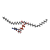


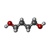


| #2: Chemical | ChemComp-ZN /  Mass: 65.409 Da / Num. of mol.: 4 / Source method: obtained synthetically / Formula: Zn Mass: 65.409 Da / Num. of mol.: 4 / Source method: obtained synthetically / Formula: Zn#3: Chemical |  Mass: 1006.147 Da / Num. of mol.: 2 / Source method: obtained synthetically / Formula: C48H85N3O15P2 / Feature type: SUBJECT OF INVESTIGATION Mass: 1006.147 Da / Num. of mol.: 2 / Source method: obtained synthetically / Formula: C48H85N3O15P2 / Feature type: SUBJECT OF INVESTIGATION#4: Chemical | ChemComp-G3P /  Mass: 172.074 Da / Num. of mol.: 5 / Source method: obtained synthetically / Formula: C3H9O6P Mass: 172.074 Da / Num. of mol.: 5 / Source method: obtained synthetically / Formula: C3H9O6P#5: Chemical |  Mass: 356.540 Da / Num. of mol.: 3 / Source method: isolated from a natural source / Formula: C21H40O4 Mass: 356.540 Da / Num. of mol.: 3 / Source method: isolated from a natural source / Formula: C21H40O4#6: Chemical | ChemComp-BU1 /  Mass: 90.121 Da / Num. of mol.: 10 / Source method: obtained synthetically / Formula: C4H10O2 Mass: 90.121 Da / Num. of mol.: 10 / Source method: obtained synthetically / Formula: C4H10O2#7: Chemical |  Mass: 60.052 Da / Num. of mol.: 2 / Source method: obtained synthetically / Formula: C2H4O2 Mass: 60.052 Da / Num. of mol.: 2 / Source method: obtained synthetically / Formula: C2H4O2#8: Water | ChemComp-HOH / | Mass: 18.015 Da / Num. of mol.: 104 / Source method: isolated from a natural source / Formula: H2O |
|---|
-Details
| Has ligand of interest | Y |
|---|
-Experimental details
-Experiment
| Experiment | Method: X-RAY DIFFRACTION / Number of used crystals: 1 |
|---|
- Sample preparation
| Crystal | Density Matthews: 3.07 Å3/Da / Density % sol: 59.88 % / Description: Plate-like crystal |
|---|---|
| Crystal grow | Temperature: 293 K / Method: lipidic cubic phase / pH: 5.5 Details: 15-26% (v/v) 1,4-butanediol, 0.2M zinc acetate, 0.1M imidazole/HCl (pH 5.0-7.5) PH range: 5.0 - 7.5 |
-Data collection
| Diffraction | Mean temperature: 100 K / Ambient temp details: Liquid nitrogen cooling / Serial crystal experiment: N |
|---|---|
| Diffraction source | Source: SYNCHROTRON / Site: SSRF / Beamline: BL18U1 / Wavelength: 0.97853 Å |
| Detector | Type: DECTRIS PILATUS3 S 6M / Detector: PIXEL / Date: May 23, 2019 |
| Radiation | Monochromator: LN2-cooled DCM with Si(111) crystals / Protocol: SINGLE WAVELENGTH / Monochromatic (M) / Laue (L): M / Scattering type: x-ray |
| Radiation wavelength | Wavelength: 0.97853 Å / Relative weight: 1 |
| Reflection | Resolution: 2.89→35 Å / Num. obs: 11858 / % possible obs: 99.5 % / Redundancy: 4.6 % / Biso Wilson estimate: 58.93 Å2 / Rmerge(I) obs: 0.174 / Net I/σ(I): 5.8 |
| Reflection shell | Resolution: 2.89→3.07 Å / Redundancy: 4.7 % / Rmerge(I) obs: 1.118 / Mean I/σ(I) obs: 1.4 / Num. unique obs: 2344 / % possible all: 98.7 |
- Processing
| Software |
| |||||||||||||||||||||||||||||||||||
|---|---|---|---|---|---|---|---|---|---|---|---|---|---|---|---|---|---|---|---|---|---|---|---|---|---|---|---|---|---|---|---|---|---|---|---|---|
| Refinement | Method to determine structure: MOLECULAR REPLACEMENT Starting model: 7DRJ Resolution: 3→9.99 Å / SU ML: 0.36 / Cross valid method: FREE R-VALUE / σ(F): 1.33 / Phase error: 30.23 / Stereochemistry target values: ML
| |||||||||||||||||||||||||||||||||||
| Solvent computation | Shrinkage radii: 0.9 Å / VDW probe radii: 1.11 Å / Solvent model: FLAT BULK SOLVENT MODEL | |||||||||||||||||||||||||||||||||||
| Refinement step | Cycle: LAST / Resolution: 3→9.99 Å
| |||||||||||||||||||||||||||||||||||
| Refine LS restraints |
| |||||||||||||||||||||||||||||||||||
| LS refinement shell |
|
