 Movie
Movie Controller

[English] 日本語
 Yorodumi
- PDB-5kkg: Crystal structure of E72A mutant of ancestral protein ancMT of AD... -
+ Open data

- Basic information
| Entry | Database: PDB / ID: 5kkg | ||||||
|---|---|---|---|---|---|---|---|
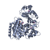
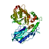
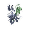
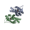
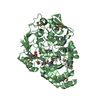
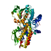
| Title | Crystal structure of E72A mutant of ancestral protein ancMT of ADP-dependent sugar kinases family | ||||||
 Components Components | ancMT E72A | ||||||
 Keywords Keywords | DE NOVO PROTEIN / ancestral protein reconstruction / ADP-dependent sugar kinases family / glucokinase / phosphofructokinase / bifuncional enzymes / enzyme evolution | ||||||
| Function / homology | Adenosine kinase, small domain - #20 / Adenosine kinase, small domain / Ribokinase / UDP-N-acetylmuramoyl-L-alanine:D-glutamate ligase / 2-Layer Sandwich / 3-Layer(aba) Sandwich / Alpha Beta / ADENOSINE MONOPHOSPHATE / IODIDE ION Function and homology information Function and homology information | ||||||
| Biological species | synthetic construct (others) | ||||||
| Method |  X-RAY DIFFRACTION / SYNCHROTRON / MOLECULAR REPLACEMENT / Resolution: 2.608 Å X-RAY DIFFRACTION / SYNCHROTRON / MOLECULAR REPLACEMENT / Resolution: 2.608 Å | ||||||
 Authors Authors | Castro-Fernandez, V. / Herrera-Morande, A. / Zamora, R. / Merino, F. / Pereira, H.M. / Brandao-Neto, J. / Garratt, R. / Guixe, V. | ||||||
| Funding support |  Chile, 1items Chile, 1items
| ||||||
 Citation Citation | Journal: J. Biol. Chem. / Year: 2017 Title: Reconstructed ancestral enzymes reveal that negative selection drove the evolution of substrate specificity in ADP-dependent kinases. Authors: Castro-Fernandez, V. / Herrera-Morande, A. / Zamora, R. / Merino, F. / Gonzalez-Ordenes, F. / Padilla-Salinas, F. / Pereira, H.M. / Brandao-Neto, J. / Garratt, R.C. / Guixe, V. | ||||||
| History |
|
- Structure visualization
| Structure viewer | Molecule: MolmilJmol/JSmol |
|---|
- Downloads & links
-Download
| PDBx/mmCIF format | 5kkg.cif.gz | 359.1 KB | Display | PDBx/mmCIF format |
|---|---|---|---|---|
| PDB format | pdb5kkg.ent.gz | 289.6 KB | Display | PDB format |
| PDBx/mmJSON format | 5kkg.json.gz | Tree view | PDBx/mmJSON format | |
| Others |  Other downloads Other downloads |
-Validation report
| Arichive directory | https://data.pdbj.org/pub/pdb/validation_reports/kk/5kkgftp://data.pdbj.org/pub/pdb/validation_reports/kk/5kkg | HTTPS FTP |
|---|
-Related structure data
| Related structure data |  5k27SC S: Starting model for refinement C: citing same article ( |
|---|---|
| Similar structure data |


-Links
 PDBj
PDBj


- Assembly
| Deposited unit | 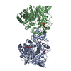
| ||||||||
|---|---|---|---|---|---|---|---|---|---|
| 1 | 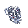
| ||||||||
| 2 | 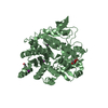
| ||||||||
| Unit cell |
|
-Components
| #1: Protein | Mass: 54255.164 Da / Num. of mol.: 2 Source method: isolated from a genetically manipulated source Source: (gene. exp.) synthetic construct (others) / Plasmid: pET-28a / Details (production host): TEV clivage site / Production host:  #2: Chemical | ChemComp-AMP / |   Mass: 347.221 Da / Num. of mol.: 1 / Source method: obtained synthetically / Formula: C10H14N5O7P / Comment: AMP*YM Mass: 347.221 Da / Num. of mol.: 1 / Source method: obtained synthetically / Formula: C10H14N5O7P / Comment: AMP*YM#3: Chemical | ChemComp-IOD /   Mass: 126.904 Da / Num. of mol.: 12 / Source method: obtained synthetically / Formula: I Mass: 126.904 Da / Num. of mol.: 12 / Source method: obtained synthetically / Formula: I#4: Chemical |   Mass: 92.094 Da / Num. of mol.: 3 / Source method: obtained synthetically / Formula: C3H8O3 Mass: 92.094 Da / Num. of mol.: 3 / Source method: obtained synthetically / Formula: C3H8O3#5: Water | ChemComp-HOH / |  Mass: 18.015 Da / Num. of mol.: 101 / Source method: isolated from a natural source / Formula: H2O Mass: 18.015 Da / Num. of mol.: 101 / Source method: isolated from a natural source / Formula: H2O |
|---|
-Experimental details
-Experiment
| Experiment | Method: X-RAY DIFFRACTION / Number of used crystals: 1 |
|---|
- Sample preparation
| Crystal | Density Matthews: 2.48 Å3/Da / Density % sol: 50.36 % |
|---|---|
| Crystal grow | Temperature: 291 K / Method: vapor diffusion, hanging drop / pH: 7.8 Details: 18 % (w/v) PEG 3350, 0.2 M NaI Protein 8 mg/mL, 20 mM F6P, 30 mM MgCl2, 25 mM HEPES pH 7.8, 20 mM AMP |
-Data collection
| Diffraction | Mean temperature: 100 K | |||||||||||||||
|---|---|---|---|---|---|---|---|---|---|---|---|---|---|---|---|---|
| Diffraction source | Source: SYNCHROTRON / Site: Diamond  / Beamline: I04-1 / Wavelength: 0.92 Å / Beamline: I04-1 / Wavelength: 0.92 Å | |||||||||||||||
| Detector | Type: DECTRIS PILATUS 2M / Detector: PIXEL / Date: Apr 26, 2014 | |||||||||||||||
| Radiation | Protocol: SINGLE WAVELENGTH / Monochromatic (M) / Laue (L): M / Scattering type: x-ray | |||||||||||||||
| Radiation wavelength | Wavelength: 0.92 Å / Relative weight: 1 | |||||||||||||||
| Reflection | Resolution: 2.608→59.27 Å / Num. obs: 31763 / % possible obs: 97.7 % / Redundancy: 8.5 % / Biso Wilson estimate: 56.06 Å2 / CC1/2: 0.995 / Rmerge(I) obs: 0.122 / Net I/σ(I): 11.8 | |||||||||||||||
| Reflection shell |
|
- Processing
| Software |
| ||||||||||||||||||||||||||||||||||||||||||||||||||||||||||||||||||||||||||||||||||||||||||||||||||||||||||||||||||||||||||||||||||||||||||||||||||||||
|---|---|---|---|---|---|---|---|---|---|---|---|---|---|---|---|---|---|---|---|---|---|---|---|---|---|---|---|---|---|---|---|---|---|---|---|---|---|---|---|---|---|---|---|---|---|---|---|---|---|---|---|---|---|---|---|---|---|---|---|---|---|---|---|---|---|---|---|---|---|---|---|---|---|---|---|---|---|---|---|---|---|---|---|---|---|---|---|---|---|---|---|---|---|---|---|---|---|---|---|---|---|---|---|---|---|---|---|---|---|---|---|---|---|---|---|---|---|---|---|---|---|---|---|---|---|---|---|---|---|---|---|---|---|---|---|---|---|---|---|---|---|---|---|---|---|---|---|---|---|---|---|
| Refinement | Method to determine structure: MOLECULAR REPLACEMENT Starting model: 5K27 Resolution: 2.608→59.269 Å / SU ML: 0.33 / Cross valid method: FREE R-VALUE / σ(F): 1.34 / Phase error: 26.4
| ||||||||||||||||||||||||||||||||||||||||||||||||||||||||||||||||||||||||||||||||||||||||||||||||||||||||||||||||||||||||||||||||||||||||||||||||||||||
| Solvent computation | Shrinkage radii: 0.9 Å / VDW probe radii: 1.11 Å | ||||||||||||||||||||||||||||||||||||||||||||||||||||||||||||||||||||||||||||||||||||||||||||||||||||||||||||||||||||||||||||||||||||||||||||||||||||||
| Displacement parameters | Biso max: 142.35 Å2 / Biso mean: 64.8791 Å2 / Biso min: 23.24 Å2 | ||||||||||||||||||||||||||||||||||||||||||||||||||||||||||||||||||||||||||||||||||||||||||||||||||||||||||||||||||||||||||||||||||||||||||||||||||||||
| Refinement step | Cycle: final / Resolution: 2.608→59.269 Å
| ||||||||||||||||||||||||||||||||||||||||||||||||||||||||||||||||||||||||||||||||||||||||||||||||||||||||||||||||||||||||||||||||||||||||||||||||||||||
| Refine LS restraints |
| ||||||||||||||||||||||||||||||||||||||||||||||||||||||||||||||||||||||||||||||||||||||||||||||||||||||||||||||||||||||||||||||||||||||||||||||||||||||
| LS refinement shell | Refine-ID: X-RAY DIFFRACTION / Total num. of bins used: 11
| ||||||||||||||||||||||||||||||||||||||||||||||||||||||||||||||||||||||||||||||||||||||||||||||||||||||||||||||||||||||||||||||||||||||||||||||||||||||
| Refinement TLS params. | Method: refined / Refine-ID: X-RAY DIFFRACTION
| ||||||||||||||||||||||||||||||||||||||||||||||||||||||||||||||||||||||||||||||||||||||||||||||||||||||||||||||||||||||||||||||||||||||||||||||||||||||
| Refinement TLS group |
|
