 Movie
Movie Controller


+ Open data

- Basic information
| Entry | Database: PDB / ID: 1pq9 | ||||||
|---|---|---|---|---|---|---|---|
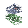
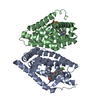
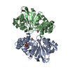
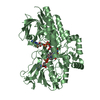
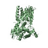
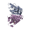
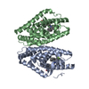
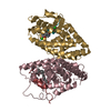
| Title | HUMAN LXR BETA HORMONE RECEPTOR COMPLEXED WITH T0901317 COMPLEX | ||||||
 Components Components | Oxysterols receptor LXR-beta | ||||||
 Keywords Keywords | transcription regulation / LXRB+T0901317 SPLIT | ||||||
| Function / homology |  Function and homology information Function and homology informationpositive regulation of secretion of lysosomal enzymes / positive regulation of high-density lipoprotein particle assembly / positive regulation of pancreatic juice secretion / phosphatidylcholine acyl-chain remodeling / negative regulation of response to endoplasmic reticulum stress / negative regulation of pinocytosis / regulation of lipid storage / positive regulation of triglyceride biosynthetic process / apolipoprotein A-I receptor binding / NR1H2 & NR1H3 regulate gene expression linked to triglyceride lipolysis in adipose ...positive regulation of secretion of lysosomal enzymes / positive regulation of high-density lipoprotein particle assembly / positive regulation of pancreatic juice secretion / phosphatidylcholine acyl-chain remodeling / negative regulation of response to endoplasmic reticulum stress / negative regulation of pinocytosis / regulation of lipid storage / positive regulation of triglyceride biosynthetic process / apolipoprotein A-I receptor binding / NR1H2 & NR1H3 regulate gene expression linked to triglyceride lipolysis in adipose / NR1H2 & NR1H3 regulate gene expression to limit cholesterol uptake / NR1H2 & NR1H3 regulate gene expression linked to lipogenesis / positive regulation of fatty acid biosynthetic process / positive regulation of lipid storage / negative regulation of lipid transport / negative regulation of cold-induced thermogenesis / positive regulation of cholesterol transport / NR1H2 & NR1H3 regulate gene expression to control bile acid homeostasis / negative regulation of type II interferon-mediated signaling pathway / negative regulation of cholesterol storage / negative regulation of macrophage derived foam cell differentiation / positive regulation of cholesterol efflux / nuclear retinoid X receptor binding / retinoic acid receptor signaling pathway / NR1H3 & NR1H2 regulate gene expression linked to cholesterol transport and efflux / intracellular receptor signaling pathway / hormone-mediated signaling pathway / negative regulation of proteolysis / cholesterol homeostasis / VLDLR internalisation and degradation / SUMOylation of intracellular receptors / PPARA activates gene expression / Nuclear Receptor transcription pathway / chromatin DNA binding / response to nutrient levels / negative regulation of inflammatory response / mRNA transcription by RNA polymerase II / nuclear receptor activity / RNA polymerase II transcription regulator complex / ATPase binding / DNA-binding transcription activator activity, RNA polymerase II-specific / DNA-binding transcription factor activity, RNA polymerase II-specific / cell differentiation / RNA polymerase II cis-regulatory region sequence-specific DNA binding / negative regulation of gene expression / negative regulation of DNA-templated transcription / positive regulation of gene expression / positive regulation of DNA-templated transcription / chromatin / negative regulation of transcription by RNA polymerase II / positive regulation of transcription by RNA polymerase II / DNA binding / zinc ion binding / nucleoplasm / nucleus / cytosol / cytoplasm Similarity search - Function | ||||||
| Biological species |  Homo sapiens (human) Homo sapiens (human) | ||||||
| Method |  X-RAY DIFFRACTION / SYNCHROTRON / MOLECULAR REPLACEMENT / Resolution: 2.1 Å X-RAY DIFFRACTION / SYNCHROTRON / MOLECULAR REPLACEMENT / Resolution: 2.1 Å | ||||||
 Authors Authors | Farnegardh, M. / Bonn, T. / Sun, S. / Ljunggren, J. / Ahola, H. / Wilhelmsson, A. / Gustafsson, J.-A. / Carlquist, M. | ||||||
 Citation Citation | Journal: J.Biol.Chem. / Year: 2003 Title: The three-dimensional structure of the liver X receptor beta reveals a flexible ligand-binding pocket that can accommodate fundamentally different ligands. Authors: Farnegardh, M. / Bonn, T. / Sun, S. / Ljunggren, J. / Ahola, H. / Wilhelmsson, A. / Gustafsson, J.-A. / Carlquist, M. | ||||||
| History |
|
- Structure visualization
| Structure viewer | Molecule: MolmilJmol/JSmol |
|---|
- Downloads & links
-Download
| PDBx/mmCIF format | 1pq9.cif.gz | 196.7 KB | Display | PDBx/mmCIF format |
|---|---|---|---|---|
| PDB format | pdb1pq9.ent.gz | 159.3 KB | Display | PDB format |
| PDBx/mmJSON format | 1pq9.json.gz | Tree view | PDBx/mmJSON format | |
| Others |  Other downloads Other downloads |
-Validation report
| Arichive directory | https://data.pdbj.org/pub/pdb/validation_reports/pq/1pq9ftp://data.pdbj.org/pub/pdb/validation_reports/pq/1pq9 | HTTPS FTP |
|---|
-Related structure data


-Links
 PDBj
PDBj


- Assembly
| Deposited unit | 
| ||||||||
|---|---|---|---|---|---|---|---|---|---|
| 1 | 
| ||||||||
| 2 | 
| ||||||||
| 3 | 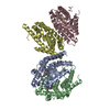
| ||||||||
| 4 | 
| ||||||||
| Unit cell |
|
-Components
| #1: Protein | Mass: 29057.283 Da / Num. of mol.: 4 / Fragment: Ligand binding domain, residues 213-461 Source method: isolated from a genetically manipulated source Source: (gene. exp.) Homo sapiens (human) / Gene: NR1H2 OR LXRB OR UNR OR NER / Plasmid: pet 28a / Production host:  #2: Chemical | ChemComp-BNS /   Mass: 158.175 Da / Num. of mol.: 4 / Source method: obtained synthetically / Formula: C6H6O3S Mass: 158.175 Da / Num. of mol.: 4 / Source method: obtained synthetically / Formula: C6H6O3S#3: Chemical | ChemComp-44B /   Mass: 341.173 Da / Num. of mol.: 4 / Source method: obtained synthetically / Formula: C11H8F9NO Mass: 341.173 Da / Num. of mol.: 4 / Source method: obtained synthetically / Formula: C11H8F9NO#4: Water | ChemComp-HOH / |  Mass: 18.015 Da / Num. of mol.: 181 / Source method: isolated from a natural source / Formula: H2O Mass: 18.015 Da / Num. of mol.: 181 / Source method: isolated from a natural source / Formula: H2O |
|---|
-Experimental details
-Experiment
| Experiment | Method: X-RAY DIFFRACTION / Number of used crystals: 1 |
|---|
- Sample preparation
| Crystal | Density Matthews: 2.31 Å3/Da / Density % sol: 46.82 % | ||||||||||||||||||||||||||||||
|---|---|---|---|---|---|---|---|---|---|---|---|---|---|---|---|---|---|---|---|---|---|---|---|---|---|---|---|---|---|---|---|
| Crystal grow | Temperature: 292 K / Method: vapor diffusion, hanging drop / pH: 7.5 Details: isopropanol, peg 4000, hepes, glycerol, pH 7.5, VAPOR DIFFUSION, HANGING DROP, temperature 292K | ||||||||||||||||||||||||||||||
| Crystal grow | *PLUS Method: vapor diffusion, hanging drop | ||||||||||||||||||||||||||||||
| Components of the solutions | *PLUS
|
-Data collection
| Diffraction | Mean temperature: 100 K |
|---|---|
| Diffraction source | Source: SYNCHROTRON / Site: ESRF  / Beamline: ID14-4 / Wavelength: 0.9393 Å / Beamline: ID14-4 / Wavelength: 0.9393 Å |
| Detector | Type: ADSC QUANTUM 4 / Detector: CCD / Date: Feb 22, 2002 / Details: toroidal mirrors |
| Radiation | Monochromator: Double crystal, Si(111) / Protocol: SINGLE WAVELENGTH / Monochromatic (M) / Laue (L): M / Scattering type: x-ray |
| Radiation wavelength | Wavelength: 0.9393 Å / Relative weight: 1 |
| Reflection | Resolution: 2.1→55.84 Å / Num. all: 63700 / Num. obs: 63700 / % possible obs: 100 % / Observed criterion σ(I): 0 / Redundancy: 3.7 % / Biso Wilson estimate: 35.44 Å2 / Rsym value: 0.276 / Net I/σ(I): 5 |
| Reflection shell | Resolution: 2.1→2.155 Å / Redundancy: 3.7 % / Mean I/σ(I) obs: 2.7 / Num. unique all: 9212 / Rsym value: 0.066 / % possible all: 100 |
| Reflection | *PLUS Num. obs: 62281 / % possible obs: 99.8 % / Num. measured all: 231983 / Rmerge(I) obs: 0.066 |
| Reflection shell | *PLUS Lowest resolution: 2.21 Å / % possible obs: 100 % / Rmerge(I) obs: 0.276 |
- Processing
| Software |
| ||||||||||||||||||||||||||||||||||||||||||||||||||||||||||||||||||||||||||||||||||||||||||||||||||||
|---|---|---|---|---|---|---|---|---|---|---|---|---|---|---|---|---|---|---|---|---|---|---|---|---|---|---|---|---|---|---|---|---|---|---|---|---|---|---|---|---|---|---|---|---|---|---|---|---|---|---|---|---|---|---|---|---|---|---|---|---|---|---|---|---|---|---|---|---|---|---|---|---|---|---|---|---|---|---|---|---|---|---|---|---|---|---|---|---|---|---|---|---|---|---|---|---|---|---|---|---|---|
| Refinement | Method to determine structure: MOLECULAR REPLACEMENT Starting model: Thyroid hormone receptor beta ligand binding domain Resolution: 2.1→55.84 Å / Cor.coef. Fo:Fc: 0.941 / Cor.coef. Fo:Fc free: 0.924 / SU B: 5.4 / SU ML: 0.145 / Isotropic thermal model: Isotropic / Cross valid method: THROUGHOUT / σ(F): 0 / ESU R: 0.236 / ESU R Free: 0.195 / Stereochemistry target values: MAXIMUM LIKELIHOOD / Details: HYDROGENS HAVE BEEN ADDED IN THE RIDING POSITIONS
| ||||||||||||||||||||||||||||||||||||||||||||||||||||||||||||||||||||||||||||||||||||||||||||||||||||
| Solvent computation | Ion probe radii: 0.8 Å / Shrinkage radii: 0.8 Å / VDW probe radii: 1.4 Å / Solvent model: BABINET MODEL WITH MASK | ||||||||||||||||||||||||||||||||||||||||||||||||||||||||||||||||||||||||||||||||||||||||||||||||||||
| Displacement parameters | Biso mean: 20.228 Å2
| ||||||||||||||||||||||||||||||||||||||||||||||||||||||||||||||||||||||||||||||||||||||||||||||||||||
| Refinement step | Cycle: LAST / Resolution: 2.1→55.84 Å
| ||||||||||||||||||||||||||||||||||||||||||||||||||||||||||||||||||||||||||||||||||||||||||||||||||||
| Refine LS restraints |
| ||||||||||||||||||||||||||||||||||||||||||||||||||||||||||||||||||||||||||||||||||||||||||||||||||||
| LS refinement shell | Resolution: 2.1→2.155 Å / Total num. of bins used: 20 /
| ||||||||||||||||||||||||||||||||||||||||||||||||||||||||||||||||||||||||||||||||||||||||||||||||||||
| Refinement | *PLUS Highest resolution: 2.1 Å / Rfactor Rfree: 0.279 / Rfactor Rwork: 0.243 | ||||||||||||||||||||||||||||||||||||||||||||||||||||||||||||||||||||||||||||||||||||||||||||||||||||
| Solvent computation | *PLUS | ||||||||||||||||||||||||||||||||||||||||||||||||||||||||||||||||||||||||||||||||||||||||||||||||||||
| Displacement parameters | *PLUS | ||||||||||||||||||||||||||||||||||||||||||||||||||||||||||||||||||||||||||||||||||||||||||||||||||||
| Refine LS restraints | *PLUS
| ||||||||||||||||||||||||||||||||||||||||||||||||||||||||||||||||||||||||||||||||||||||||||||||||||||
| LS refinement shell | *PLUS Highest resolution: 2.1 Å |
