 Movie
Movie Controller

[English] 日本語
 Yorodumi
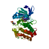
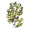
- PDB-7dqn: The structure of the Arabidopsis thaliana guanosine deaminase mut... -
+ Open data

- Basic information
| Entry | Database: PDB / ID: 7dqn | ||||||
|---|---|---|---|---|---|---|---|
| Title | The structure of the Arabidopsis thaliana guanosine deaminase mutant Y185F complexed with guanosine | ||||||
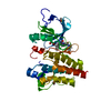
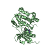
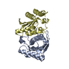
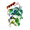
 Components Components | Guanosine deaminase | ||||||
 Keywords Keywords | HYDROLASE / deamination / GSDA / purine metabolism / plant protein | ||||||
| Function / homology |  Function and homology information Function and homology informationguanosine deaminase / guanosine deaminase activity / purine nucleoside catabolic process / plastid / zinc ion binding / nucleus / cytoplasm / cytosol Similarity search - Function | ||||||
| Biological species |  | ||||||
| Method |  X-RAY DIFFRACTION / MOLECULAR REPLACEMENT / Resolution: 2.6 Å X-RAY DIFFRACTION / MOLECULAR REPLACEMENT / Resolution: 2.6 Å | ||||||
 Authors Authors | Xie, W. / Jia, Q. / Zeng, H. | ||||||
| Funding support |  China, 1items China, 1items
| ||||||
 Citation Citation | Journal: Int J Mol Sci / Year: 2022 Title: Substrate Specificity of GSDA Revealed by Cocrystal Structures and Binding Studies. Authors: Jia, Q. / Zhang, J. / Zeng, H. / Tang, J. / Xiao, N. / Gao, S. / Li, H. / Xie, W. | ||||||
| History |
|
- Structure visualization
| Structure viewer | Molecule: MolmilJmol/JSmol |
|---|
- Downloads & links
-Download
| PDBx/mmCIF format | 7dqn.cif.gz | 90 KB | Display | PDBx/mmCIF format |
|---|---|---|---|---|
| PDB format | pdb7dqn.ent.gz | 54.6 KB | Display | PDB format |
| PDBx/mmJSON format | 7dqn.json.gz | Tree view | PDBx/mmJSON format | |
| Others |  Other downloads Other downloads |
-Validation report
| Arichive directory | https://data.pdbj.org/pub/pdb/validation_reports/dq/7dqnftp://data.pdbj.org/pub/pdb/validation_reports/dq/7dqn | HTTPS FTP |
|---|
-Related structure data
| Related structure data |  7dcbC  7dcwC 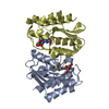 7dgcC  7dh1C  7dm5C  7dm6C 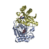 7w1qC  7dbfS S: Starting model for refinement C: citing same article ( |
|---|---|
| Similar structure data |

-Links
 PDBj
PDBj- Assembly
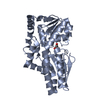
| Deposited unit | 
| ||||||||||||
|---|---|---|---|---|---|---|---|---|---|---|---|---|---|
| 1 |
| ||||||||||||
| Unit cell |
|
-Components
| #1: Protein | Mass: 17507.016 Da / Num. of mol.: 2 / Mutation: Y185F Source method: isolated from a genetically manipulated source Source: (gene. exp.)  #2: Chemical | ChemComp-GMP / | 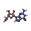  Mass: 283.241 Da / Num. of mol.: 1 / Source method: obtained synthetically / Formula: C10H13N5O5 / Feature type: SUBJECT OF INVESTIGATION Mass: 283.241 Da / Num. of mol.: 1 / Source method: obtained synthetically / Formula: C10H13N5O5 / Feature type: SUBJECT OF INVESTIGATION#3: Chemical |   Mass: 65.409 Da / Num. of mol.: 2 / Source method: isolated from a natural source / Formula: Zn / Feature type: SUBJECT OF INVESTIGATION Mass: 65.409 Da / Num. of mol.: 2 / Source method: isolated from a natural source / Formula: Zn / Feature type: SUBJECT OF INVESTIGATION#4: Water | ChemComp-HOH / |  Mass: 18.015 Da / Num. of mol.: 92 / Source method: isolated from a natural source / Formula: H2O Mass: 18.015 Da / Num. of mol.: 92 / Source method: isolated from a natural source / Formula: H2OHas ligand of interest | Y | |
|---|
-Experimental details
-Experiment
| Experiment | Method: X-RAY DIFFRACTION / Number of used crystals: 1 |
|---|
- Sample preparation
| Crystal | Density Matthews: 2.31 Å3/Da / Density % sol: 46.78 % |
|---|---|
| Crystal grow | Temperature: 277 K / Method: vapor diffusion, sitting drop / Details: 1.2 M Na-citrate and 0.1 M HEPES (pH 7.5) |
-Data collection
| Diffraction | Mean temperature: 100 K / Serial crystal experiment: N |
|---|---|
| Diffraction source | Source: ROTATING ANODE / Type: RIGAKU MICROMAX-007 HF / Wavelength: 1.54 Å |
| Detector | Type: OXFORD ONYX CCD / Detector: CCD / Date: Dec 16, 2020 |
| Radiation | Protocol: SINGLE WAVELENGTH / Monochromatic (M) / Laue (L): M / Scattering type: x-ray |
| Radiation wavelength | Wavelength: 1.54 Å / Relative weight: 1 |
| Reflection | Resolution: 2.6→23.78 Å / Num. obs: 10100 / % possible obs: 99.9 % / Redundancy: 6.9 % / Biso Wilson estimate: 25.29 Å2 / CC1/2: 0.992 / Rmerge(I) obs: 0.162 / Net I/σ(I): 12.1 |
| Reflection shell | Resolution: 2.6→2.74 Å / Redundancy: 7.1 % / Rmerge(I) obs: 0.609 / Mean I/σ(I) obs: 3.7 / Num. unique obs: 1442 / CC1/2: 0.838 / % possible all: 100 |
- Processing
| Software |
| ||||||||||||||||||||||||||||
|---|---|---|---|---|---|---|---|---|---|---|---|---|---|---|---|---|---|---|---|---|---|---|---|---|---|---|---|---|---|
| Refinement | Method to determine structure: MOLECULAR REPLACEMENT Starting model: 7DBF Resolution: 2.6→23.78 Å / SU ML: 0.2894 / Cross valid method: FREE R-VALUE / σ(F): 1.36 / Phase error: 22.2307 / Stereochemistry target values: CDL v1.2
| ||||||||||||||||||||||||||||
| Solvent computation | Shrinkage radii: 0.9 Å / VDW probe radii: 1.11 Å / Solvent model: FLAT BULK SOLVENT MODEL | ||||||||||||||||||||||||||||
| Displacement parameters | Biso mean: 27.62 Å2 | ||||||||||||||||||||||||||||
| Refinement step | Cycle: LAST / Resolution: 2.6→23.78 Å
| ||||||||||||||||||||||||||||
| Refine LS restraints |
| ||||||||||||||||||||||||||||
| LS refinement shell |
|
