 Movie
Movie Controller


+ Open data

- Basic information
| Entry | Database: PDB / ID: 6jil | ||||||
|---|---|---|---|---|---|---|---|
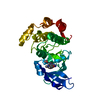
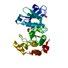
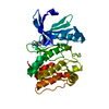
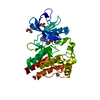
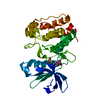
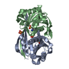
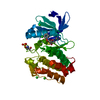
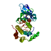
| Title | Crystal structure of D-cycloserine synthetase DcsG | ||||||
 Components Components | Cycloserine biosynthesis protein DcsG | ||||||
 Keywords Keywords | LIGASE / ATP-grasp family / antibiotic / biosynthesis | ||||||
| Function / homology |  Function and homology information Function and homology informationO-ureido-D-serine cyclo-ligase / cyclo-ligase activity / antibiotic biosynthetic process / ATP binding / metal ion binding Similarity search - Function | ||||||
| Biological species |  Streptomyces lavendulae (bacteria) Streptomyces lavendulae (bacteria) | ||||||
| Method |  X-RAY DIFFRACTION / SYNCHROTRON / SAD / Resolution: 2.32 Å X-RAY DIFFRACTION / SYNCHROTRON / SAD / Resolution: 2.32 Å | ||||||
 Authors Authors | Matoba, Y. / Sugiyama, M. | ||||||
| Funding support |  Japan, 1items Japan, 1items
| ||||||
 Citation Citation | Journal: Febs J. / Year: 2020 Title: Cyclization mechanism catalyzed by an ATP-grasp enzyme essential for d-cycloserine biosynthesis. Authors: Matoba, Y. / Uda, N. / Kudo, M. / Sugiyama, M. | ||||||
| History |
|
- Structure visualization
| Structure viewer | Molecule: MolmilJmol/JSmol |
|---|
- Downloads & links
-Download
| PDBx/mmCIF format | 6jil.cif.gz | 261.8 KB | Display | PDBx/mmCIF format |
|---|---|---|---|---|
| PDB format | pdb6jil.ent.gz | 210.8 KB | Display | PDB format |
| PDBx/mmJSON format | 6jil.json.gz | Tree view | PDBx/mmJSON format | |
| Others |  Other downloads Other downloads |
-Validation report
| Arichive directory | https://data.pdbj.org/pub/pdb/validation_reports/ji/6jilftp://data.pdbj.org/pub/pdb/validation_reports/ji/6jil | HTTPS FTP |
|---|
-Related structure data
| Similar structure data |
|---|

-Links
 PDBj
PDBj


- Assembly
| Deposited unit | 
| ||||||||
|---|---|---|---|---|---|---|---|---|---|
| 1 | 
| ||||||||
| 2 | 
| ||||||||
| 3 | 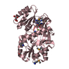
| ||||||||
| 4 | 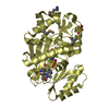
| ||||||||
| Unit cell |
|
-Components
| #1: Protein | Mass: 34222.836 Da / Num. of mol.: 4 Source method: isolated from a genetically manipulated source Source: (gene. exp.) Streptomyces lavendulae (bacteria) / Gene: dcsG / Plasmid: pET-dcsG / Production host: #2: Chemical | ChemComp-TLA /   Mass: 150.087 Da / Num. of mol.: 10 / Source method: obtained synthetically / Formula: C4H6O6 Mass: 150.087 Da / Num. of mol.: 10 / Source method: obtained synthetically / Formula: C4H6O6#3: Chemical | ChemComp-ADP /   Mass: 427.201 Da / Num. of mol.: 4 / Source method: obtained synthetically / Formula: C10H15N5O10P2 / Comment: ADP, energy-carrying molecule*YM Mass: 427.201 Da / Num. of mol.: 4 / Source method: obtained synthetically / Formula: C10H15N5O10P2 / Comment: ADP, energy-carrying molecule*YM#4: Chemical | ChemComp-MG /   Mass: 24.305 Da / Num. of mol.: 9 / Source method: obtained synthetically / Formula: Mg Mass: 24.305 Da / Num. of mol.: 9 / Source method: obtained synthetically / Formula: Mg#5: Water | ChemComp-HOH / |  Mass: 18.015 Da / Num. of mol.: 756 / Source method: isolated from a natural source / Formula: H2O Mass: 18.015 Da / Num. of mol.: 756 / Source method: isolated from a natural source / Formula: H2OHas protein modification | Y | |
|---|
-Experimental details
-Experiment
| Experiment | Method: X-RAY DIFFRACTION / Number of used crystals: 1 |
|---|
- Sample preparation
| Crystal | Density Matthews: 2.51 Å3/Da / Density % sol: 51.04 % |
|---|---|
| Crystal grow | Temperature: 298 K / Method: vapor diffusion, sitting drop / pH: 7.5 Details: 1.0 M potassium sodium L-tartrate, 0.1 M Tris-HCl buffer (pH 7.5) |
-Data collection
| Diffraction | Mean temperature: 100 K / Serial crystal experiment: N |
|---|---|
| Diffraction source | Source: SYNCHROTRON / Site: SPring-8 / Beamline: BL38B1 / Wavelength: 0.97864 Å |
| Detector | Type: RAYONIX MX-225 / Detector: CCD / Date: Jan 20, 2015 |
| Radiation | Protocol: SINGLE WAVELENGTH / Monochromatic (M) / Laue (L): M / Scattering type: x-ray |
| Radiation wavelength | Wavelength: 0.97864 Å / Relative weight: 1 |
| Reflection | Resolution: 2.32→100 Å / Num. obs: 55347 / % possible obs: 95.3 % / Redundancy: 3.6 % / Biso Wilson estimate: 20.1 Å2 / Rmerge(I) obs: 0.117 / Net I/σ(I): 10.1 |
| Reflection shell | Resolution: 2.32→2.4 Å / Redundancy: 2.9 % / Rmerge(I) obs: 0.447 / Mean I/σ(I) obs: 1.8 / % possible all: 82.4 |
- Processing
| Software |
| ||||||||||||||||||||||||||||||||||||||||||||||||||||||||||||||||||||||||||||||||
|---|---|---|---|---|---|---|---|---|---|---|---|---|---|---|---|---|---|---|---|---|---|---|---|---|---|---|---|---|---|---|---|---|---|---|---|---|---|---|---|---|---|---|---|---|---|---|---|---|---|---|---|---|---|---|---|---|---|---|---|---|---|---|---|---|---|---|---|---|---|---|---|---|---|---|---|---|---|---|---|---|---|
| Refinement | Method to determine structure: SAD / Resolution: 2.32→29.76 Å / Rfactor Rfree error: 0.005 / Data cutoff high absF: 1851637.9 / Data cutoff low absF: 0 / Cross valid method: THROUGHOUT / σ(F): 2 Details: SF FILE CONTAINS FRIEDEL PAIRS UNDER I/F_MINUS AND I/F_PLUS COLUMNS. BULK SOLVENT MODEL USED
| ||||||||||||||||||||||||||||||||||||||||||||||||||||||||||||||||||||||||||||||||
| Solvent computation | Bsol: 41.36 Å2 / ksol: 0.4 e/Å3 | ||||||||||||||||||||||||||||||||||||||||||||||||||||||||||||||||||||||||||||||||
| Displacement parameters | Biso mean: 25.1 Å2
| ||||||||||||||||||||||||||||||||||||||||||||||||||||||||||||||||||||||||||||||||
| Refine analyze |
| ||||||||||||||||||||||||||||||||||||||||||||||||||||||||||||||||||||||||||||||||
| Refinement step | Cycle: LAST / Resolution: 2.32→29.76 Å
| ||||||||||||||||||||||||||||||||||||||||||||||||||||||||||||||||||||||||||||||||
| Refine LS restraints |
| ||||||||||||||||||||||||||||||||||||||||||||||||||||||||||||||||||||||||||||||||
| LS refinement shell | Resolution: 2.32→2.47 Å / Rfactor Rfree error: 0.015 / Total num. of bins used: 6
| ||||||||||||||||||||||||||||||||||||||||||||||||||||||||||||||||||||||||||||||||
| Xplor file |
|
