 Movie
Movie Controller

[English] 日本語
 Yorodumi
- PDB-7dq6: Crystal structure of HitB in complex with (S)-beta-3-Br-phenylala... -
+ Open data

- Basic information
| Entry | Database: PDB / ID: 7dq6 | ||||||
|---|---|---|---|---|---|---|---|
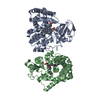
| Title | Crystal structure of HitB in complex with (S)-beta-3-Br-phenylalanine sulfamoyladenosine | ||||||
 Components Components | Putative ATP-dependent b-aminoacyl-ACP synthetase | ||||||
 Keywords Keywords | LIGASE / Hitachimycin / Polyketide biosynthesis / ATP binding / Adenylation | ||||||
| Function / homology |  Function and homology information Function and homology informationamino acid activation for nonribosomal peptide biosynthetic process / secondary metabolite biosynthetic process / phosphopantetheine binding / nucleotide binding / metal ion binding / cytoplasm Similarity search - Function | ||||||
| Biological species |  Embleya scabrispora (bacteria) Embleya scabrispora (bacteria) | ||||||
| Method |  X-RAY DIFFRACTION / SYNCHROTRON / MOLECULAR REPLACEMENT / Resolution: 2.6 Å X-RAY DIFFRACTION / SYNCHROTRON / MOLECULAR REPLACEMENT / Resolution: 2.6 Å | ||||||
 Authors Authors | Kudo, F. / Takahashi, S. / Miyanaga, A. / Nakazawa, Y. / Eguchi, T. | ||||||
 Citation Citation | Journal: Acs Chem.Biol. / Year: 2021 Title: Mutational Biosynthesis of Hitachimycin Analogs Controlled by the beta-Amino Acid-Selective Adenylation Enzyme HitB. Authors: Kudo, F. / Takahashi, S. / Miyanaga, A. / Nakazawa, Y. / Nishino, K. / Hayakawa, Y. / Kawamura, K. / Ishikawa, F. / Tanabe, G. / Iwai, N. / Nagumo, Y. / Usui, T. / Eguchi, T. | ||||||
| History |
|
- Structure visualization
| Structure viewer | Molecule: MolmilJmol/JSmol |
|---|
- Downloads & links
-Download
| PDBx/mmCIF format | 7dq6.cif.gz | 363.6 KB | Display | PDBx/mmCIF format |
|---|---|---|---|---|
| PDB format | pdb7dq6.ent.gz | 295.3 KB | Display | PDB format |
| PDBx/mmJSON format | 7dq6.json.gz | Tree view | PDBx/mmJSON format | |
| Others |  Other downloads Other downloads |
-Validation report
| Arichive directory | https://data.pdbj.org/pub/pdb/validation_reports/dq/7dq6ftp://data.pdbj.org/pub/pdb/validation_reports/dq/7dq6 | HTTPS FTP |
|---|
-Related structure data
| Related structure data |  7dq5SC S: Starting model for refinement C: citing same article ( |
|---|---|
| Similar structure data |


-Links
 PDBj
PDBj
- Assembly
| Deposited unit | 
| ||||||||||||||||||
|---|---|---|---|---|---|---|---|---|---|---|---|---|---|---|---|---|---|---|---|
| 1 |
| ||||||||||||||||||
| Unit cell |
| ||||||||||||||||||
| Noncrystallographic symmetry (NCS) | NCS domain:
NCS domain segments: Component-ID: _ / Ens-ID: 1 / Beg auth comp-ID: ARG / Beg label comp-ID: ARG / End auth comp-ID: ARG / End label comp-ID: ARG / Refine code: _ / Auth seq-ID: 9 - 528 / Label seq-ID: 25 - 544
|
-Components
| #1: Protein | Mass: 59674.695 Da / Num. of mol.: 2 Source method: isolated from a genetically manipulated source Source: (gene. exp.) Embleya scabrispora (bacteria) / Gene: hitB / Production host: #2: Chemical |   Mass: 572.390 Da / Num. of mol.: 2 / Source method: obtained synthetically / Formula: C19H22BrN7O7S / Feature type: SUBJECT OF INVESTIGATION Mass: 572.390 Da / Num. of mol.: 2 / Source method: obtained synthetically / Formula: C19H22BrN7O7S / Feature type: SUBJECT OF INVESTIGATION#3: Chemical |   Mass: 40.078 Da / Num. of mol.: 2 / Source method: obtained synthetically / Formula: Ca Mass: 40.078 Da / Num. of mol.: 2 / Source method: obtained synthetically / Formula: Ca#4: Water | ChemComp-HOH / |  Mass: 18.015 Da / Num. of mol.: 62 / Source method: isolated from a natural source / Formula: H2O Mass: 18.015 Da / Num. of mol.: 62 / Source method: isolated from a natural source / Formula: H2OHas ligand of interest | Y | |
|---|
-Experimental details
-Experiment
| Experiment | Method: X-RAY DIFFRACTION / Number of used crystals: 1 |
|---|
- Sample preparation
| Crystal | Density Matthews: 2.29 Å3/Da / Density % sol: 46.31 % |
|---|---|
| Crystal grow | Temperature: 293 K / Method: vapor diffusion, sitting drop / Details: PEG 400, calcium acetate, sodium acetate |
-Data collection
| Diffraction | Mean temperature: 100 K / Serial crystal experiment: N |
|---|---|
| Diffraction source | Source: SYNCHROTRON / Site: Photon Factory  / Beamline: BL-5A / Wavelength: 1 Å / Beamline: BL-5A / Wavelength: 1 Å |
| Detector | Type: DECTRIS PILATUS3 S 6M / Detector: PIXEL / Date: Dec 10, 2019 |
| Radiation | Monochromator: Numerical link type Si(111) double crystal monochromator Protocol: SINGLE WAVELENGTH / Monochromatic (M) / Laue (L): M / Scattering type: x-ray |
| Radiation wavelength | Wavelength: 1 Å / Relative weight: 1 |
| Reflection | Resolution: 2.6→50 Å / Num. obs: 34560 / % possible obs: 99.9 % / Redundancy: 6.6 % / CC1/2: 0.999 / Rmerge(I) obs: 0.089 / Net I/σ(I): 17.5 |
| Reflection shell | Resolution: 2.6→2.72 Å / Redundancy: 6.4 % / Rmerge(I) obs: 0.804 / Mean I/σ(I) obs: 2.5 / Num. unique obs: 4167 / CC1/2: 0.861 / % possible all: 100 |
- Processing
| Software |
| |||||||||||||||||||||||||||||||||||||||||||||||||||||||||||||||||||||||||||
|---|---|---|---|---|---|---|---|---|---|---|---|---|---|---|---|---|---|---|---|---|---|---|---|---|---|---|---|---|---|---|---|---|---|---|---|---|---|---|---|---|---|---|---|---|---|---|---|---|---|---|---|---|---|---|---|---|---|---|---|---|---|---|---|---|---|---|---|---|---|---|---|---|---|---|---|---|
| Refinement | Method to determine structure: MOLECULAR REPLACEMENT Starting model: 7DQ5 Resolution: 2.6→48.39 Å / Cor.coef. Fo:Fc: 0.944 / Cor.coef. Fo:Fc free: 0.905 / SU B: 32.333 / SU ML: 0.31 / Cross valid method: THROUGHOUT / σ(F): 0 / ESU R: 0.734 / ESU R Free: 0.333 / Stereochemistry target values: MAXIMUM LIKELIHOOD Details: U VALUES : WITH TLS ADDED HYDROGENS HAVE BEEN ADDED IN THE RIDING POSITIONS
| |||||||||||||||||||||||||||||||||||||||||||||||||||||||||||||||||||||||||||
| Solvent computation | Ion probe radii: 0.8 Å / Shrinkage radii: 0.8 Å / VDW probe radii: 1.2 Å / Solvent model: MASK | |||||||||||||||||||||||||||||||||||||||||||||||||||||||||||||||||||||||||||
| Displacement parameters | Biso max: 125.7 Å2 / Biso mean: 63.691 Å2 / Biso min: 28 Å2
| |||||||||||||||||||||||||||||||||||||||||||||||||||||||||||||||||||||||||||
| Refinement step | Cycle: final / Resolution: 2.6→48.39 Å
| |||||||||||||||||||||||||||||||||||||||||||||||||||||||||||||||||||||||||||
| Refine LS restraints |
| |||||||||||||||||||||||||||||||||||||||||||||||||||||||||||||||||||||||||||
| Refine LS restraints NCS | Ens-ID: 1 / Number: 13129 / Refine-ID: X-RAY DIFFRACTION / Type: interatomic distance / Rms dev position: 0.09 Å / Weight position: 0.05
| |||||||||||||||||||||||||||||||||||||||||||||||||||||||||||||||||||||||||||
| LS refinement shell | Resolution: 2.6→2.668 Å / Rfactor Rfree error: 0 / Total num. of bins used: 20
| |||||||||||||||||||||||||||||||||||||||||||||||||||||||||||||||||||||||||||
| Refinement TLS params. | Method: refined / Refine-ID: X-RAY DIFFRACTION
| |||||||||||||||||||||||||||||||||||||||||||||||||||||||||||||||||||||||||||
| Refinement TLS group |
|
