 Movie
Movie Controller


+ Open data

- Basic information
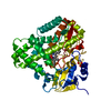
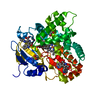
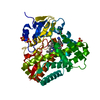
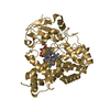
| Entry | Database: PDB / ID: 7dls | ||||||
|---|---|---|---|---|---|---|---|
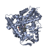
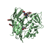
| Title | Cytochrome P450 (CYP105D18) complex with papaverine | ||||||
 Components Components | Cytochrome P450 hydroxylase | ||||||
 Keywords Keywords | TRANSFERASE / papaverine N-oxide / eukaryotic cytochrome P450 / chemical modification / heme oxidation | ||||||
| Function / homology |  Function and homology information Function and homology informationoxidoreductase activity, acting on paired donors, with incorporation or reduction of molecular oxygen / monooxygenase activity / iron ion binding / heme binding Similarity search - Function | ||||||
| Biological species |  Streptomyces laurentii (bacteria) Streptomyces laurentii (bacteria) | ||||||
| Method |  X-RAY DIFFRACTION / SYNCHROTRON / MOLECULAR REPLACEMENT / Resolution: 2.06 Å X-RAY DIFFRACTION / SYNCHROTRON / MOLECULAR REPLACEMENT / Resolution: 2.06 Å | ||||||
 Authors Authors | Do, H. / Lee, J.H. | ||||||
 Citation Citation | Journal: Iucrj / Year: 2021 Title: Characterization of high-H 2 O 2 -tolerant bacterial cytochrome P450 CYP105D18: insights into papaverine N-oxidation. Authors: Pardhe, B.D. / Do, H. / Jeong, C.S. / Kim, K.H. / Lee, J.H. / Oh, T.J. | ||||||
| History |
|
- Structure visualization
| Structure viewer | Molecule: MolmilJmol/JSmol |
|---|
- Downloads & links
-Download
| PDBx/mmCIF format | 7dls.cif.gz | 96.1 KB | Display | PDBx/mmCIF format |
|---|---|---|---|---|
| PDB format | pdb7dls.ent.gz | 69.8 KB | Display | PDB format |
| PDBx/mmJSON format | 7dls.json.gz | Tree view | PDBx/mmJSON format | |
| Others |  Other downloads Other downloads |
-Validation report
| Arichive directory | https://data.pdbj.org/pub/pdb/validation_reports/dl/7dlsftp://data.pdbj.org/pub/pdb/validation_reports/dl/7dls | HTTPS FTP |
|---|
-Related structure data
| Related structure data | 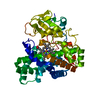 7di3SC S: Starting model for refinement C: citing same article ( |
|---|---|
| Similar structure data |


-Links
 PDBj
PDBj
- Assembly
| Deposited unit | 
| ||||||||
|---|---|---|---|---|---|---|---|---|---|
| 1 |
| ||||||||
| Unit cell |
|
-Components
| #1: Protein | Mass: 44403.266 Da / Num. of mol.: 1 Source method: isolated from a genetically manipulated source Source: (gene. exp.) Streptomyces laurentii (bacteria) / Gene: SLA_5925 / Production host: |
|---|---|
| #2: Chemical | ChemComp-HEM /   Mass: 616.487 Da / Num. of mol.: 1 / Source method: obtained synthetically / Formula: C34H32FeN4O4 / Feature type: SUBJECT OF INVESTIGATION Mass: 616.487 Da / Num. of mol.: 1 / Source method: obtained synthetically / Formula: C34H32FeN4O4 / Feature type: SUBJECT OF INVESTIGATION |
| #3: Chemical | ChemComp-EV1 / 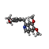  Mass: 339.385 Da / Num. of mol.: 1 / Source method: obtained synthetically / Formula: C20H21NO4 / Feature type: SUBJECT OF INVESTIGATION Mass: 339.385 Da / Num. of mol.: 1 / Source method: obtained synthetically / Formula: C20H21NO4 / Feature type: SUBJECT OF INVESTIGATION |
| #4: Water | ChemComp-HOH /  Mass: 18.015 Da / Num. of mol.: 144 / Source method: isolated from a natural source / Formula: H2O Mass: 18.015 Da / Num. of mol.: 144 / Source method: isolated from a natural source / Formula: H2O |
| Has ligand of interest | Y |
-Experimental details
-Experiment
| Experiment | Method: X-RAY DIFFRACTION / Number of used crystals: 1 |
|---|
- Sample preparation
| Crystal | Density Matthews: 2.21 Å3/Da / Density % sol: 44.36 % |
|---|---|
| Crystal grow | Temperature: 300 K / Method: vapor diffusion, sitting drop / pH: 8.5 Details: 0.1 M Tris-HCl, pH 8.5, 1.5 M Ammonium phosphate dibasic |
-Data collection
| Diffraction | Mean temperature: 100 K / Serial crystal experiment: N |
|---|---|
| Diffraction source | Source: SYNCHROTRON / Site: PAL/PLS  / Beamline: 5C (4A) / Wavelength: 0.9794 Å / Beamline: 5C (4A) / Wavelength: 0.9794 Å |
| Detector | Type: DECTRIS EIGER X 9M / Detector: PIXEL / Date: Nov 19, 2020 |
| Radiation | Protocol: SINGLE WAVELENGTH / Monochromatic (M) / Laue (L): M / Scattering type: x-ray |
| Radiation wavelength | Wavelength: 0.9794 Å / Relative weight: 1 |
| Reflection | Resolution: 2.06→50 Å / Num. obs: 41490 / % possible obs: 94.15 % / Redundancy: 4.7 % / CC1/2: 1 / Net I/σ(I): 17.05 |
| Reflection shell | Resolution: 2.06→2.1 Å / CC1/2: 1 |
- Processing
| Software |
| ||||||||||||||||||||||||||||||||||||||||||||||||||||||||||||
|---|---|---|---|---|---|---|---|---|---|---|---|---|---|---|---|---|---|---|---|---|---|---|---|---|---|---|---|---|---|---|---|---|---|---|---|---|---|---|---|---|---|---|---|---|---|---|---|---|---|---|---|---|---|---|---|---|---|---|---|---|---|
| Refinement | Method to determine structure: MOLECULAR REPLACEMENT Starting model: 7DI3 Resolution: 2.06→44.59 Å / Cor.coef. Fo:Fc: 0.959 / Cor.coef. Fo:Fc free: 0.931 / SU B: 5.128 / SU ML: 0.134 / Cross valid method: THROUGHOUT / σ(F): 0 / ESU R: 0.225 / ESU R Free: 0.181 / Stereochemistry target values: MAXIMUM LIKELIHOOD Details: HYDROGENS HAVE BEEN ADDED IN THE RIDING POSITIONS U VALUES : REFINED INDIVIDUALLY
| ||||||||||||||||||||||||||||||||||||||||||||||||||||||||||||
| Solvent computation | Ion probe radii: 0.8 Å / Shrinkage radii: 0.8 Å / VDW probe radii: 1.2 Å / Solvent model: MASK | ||||||||||||||||||||||||||||||||||||||||||||||||||||||||||||
| Displacement parameters | Biso max: 111.61 Å2 / Biso mean: 32.834 Å2 / Biso min: 14.81 Å2
| ||||||||||||||||||||||||||||||||||||||||||||||||||||||||||||
| Refinement step | Cycle: final / Resolution: 2.06→44.59 Å
| ||||||||||||||||||||||||||||||||||||||||||||||||||||||||||||
| Refine LS restraints |
| ||||||||||||||||||||||||||||||||||||||||||||||||||||||||||||
| LS refinement shell | Resolution: 2.064→2.117 Å / Rfactor Rfree error: 0 / Total num. of bins used: 20
|
