| Entry | Database: PDB / ID: 7dd0
|
|---|
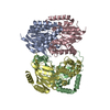
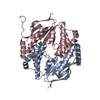
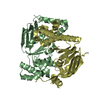
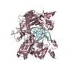
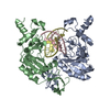
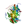
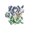
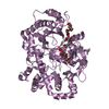
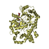
| Title | Crystal structure of the N-terminal domain of TagH from Bacillus subtilis |
|---|
 Components Components | Teichoic acids export ATP-binding protein TagH |
|---|
 Keywords Keywords | TRANSPROT PROTEIN / TRANSLOCASE / ABC transporter / ATP-binding protein / TRANSPORT PROTEIN |
|---|
| Function / homology |  Function and homology information Function and homology information
ABC-type teichoic-acid transporter / ABC-type teichoic acid transporter activity / ATP hydrolysis activity / ATP binding / plasma membraneSimilarity search - Function Teichoic acids export ATP-binding protein tagH. / ABC transporter, teichoic acids export TagH-like / : / ABC transporter-like, conserved site / ABC transporters family signature. / ABC transporter / ABC transporter-like, ATP-binding domain / ATP-binding cassette, ABC transporter-type domain profile. / P-loop containing nucleotide triphosphate hydrolases / ATPases associated with a variety of cellular activities ...Teichoic acids export ATP-binding protein tagH. / ABC transporter, teichoic acids export TagH-like / : / ABC transporter-like, conserved site / ABC transporters family signature. / ABC transporter / ABC transporter-like, ATP-binding domain / ATP-binding cassette, ABC transporter-type domain profile. / P-loop containing nucleotide triphosphate hydrolases / ATPases associated with a variety of cellular activities / AAA+ ATPase domain / Rossmann fold / P-loop containing nucleoside triphosphate hydrolase / 3-Layer(aba) Sandwich / Alpha BetaSimilarity search - Domain/homology |
|---|
| Biological species |   Bacillus subtilis subsp. subtilis str. 168 (bacteria) Bacillus subtilis subsp. subtilis str. 168 (bacteria) |
|---|
| Method |  X-RAY DIFFRACTION / SYNCHROTRON / SAD / Resolution: 2.7 Å X-RAY DIFFRACTION / SYNCHROTRON / SAD / Resolution: 2.7 Å |
|---|
 Authors Authors | Ko, T.P. / Yang, C.S. / Wang, Y.C. / Chen, Y. |
|---|
| Funding support |  Taiwan, 1items Taiwan, 1items | Organization | Grant number | Country |
|---|
| Ministry of Science and Technology (MoST, Taiwan) | 108-2311-B241-001 | Taiwan |
|
|---|
 Citation Citation | Journal: Biochem.Biophys.Res.Commun. / Year: 2020
Title: Crystal structure of the N-terminal domain of TagH reveals a potential drug targeting site.
Authors: Yang, C.S. / Huang, W.C. / Ko, T.P. / Wang, Y.C. / Wang, A.H. / Chen, Y. |
|---|
| History | | Deposition | Oct 27, 2020 | Deposition site: PDBJ / Processing site: PDBJ |
|---|
| Revision 1.0 | Dec 23, 2020 | Provider: repository / Type: Initial release |
|---|
| Revision 1.1 | Jan 13, 2021 | Group: Database references / Category: citation / citation_author
Item: _citation.country / _citation.journal_abbrev ..._citation.country / _citation.journal_abbrev / _citation.journal_id_ASTM / _citation.journal_id_CSD / _citation.journal_id_ISSN / _citation.journal_volume / _citation.page_first / _citation.page_last / _citation.pdbx_database_id_DOI / _citation.pdbx_database_id_PubMed / _citation.title / _citation.year / _citation_author.name |
|---|
| Revision 1.2 | Mar 27, 2024 | Group: Data collection / Database references ...Data collection / Database references / Derived calculations / Refinement description
Category: atom_type / chem_comp_atom ...atom_type / chem_comp_atom / chem_comp_bond / database_2 / struct_ncs_dom_lim
Item: _atom_type.pdbx_N_electrons / _atom_type.pdbx_scat_Z ..._atom_type.pdbx_N_electrons / _atom_type.pdbx_scat_Z / _database_2.pdbx_DOI / _database_2.pdbx_database_accession / _struct_ncs_dom_lim.beg_auth_comp_id / _struct_ncs_dom_lim.beg_label_asym_id / _struct_ncs_dom_lim.beg_label_comp_id / _struct_ncs_dom_lim.beg_label_seq_id / _struct_ncs_dom_lim.end_auth_comp_id / _struct_ncs_dom_lim.end_label_asym_id / _struct_ncs_dom_lim.end_label_comp_id / _struct_ncs_dom_lim.end_label_seq_id |
|---|
|
|---|
 Movie
Movie Controller

 Yorodumi
 Open data
 Basic information
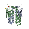
 Structure visualization
 Downloads & links
 Other downloads
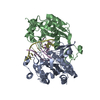

 PDBj
PDBj

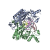
 Assembly