 Movie
Movie Controller

[English] 日本語
 Yorodumi
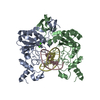
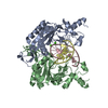
- PDB-1bua: STRUCTURAL AND ENERGETIC ORIGINS OF INDIRECT READOUT IN SITE-SPEC... -
+ Open data

- Basic information
| Entry | Database: PDB / ID: 1bua | ||||||
|---|---|---|---|---|---|---|---|
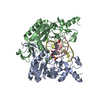
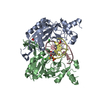
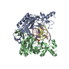
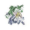
| Title | STRUCTURAL AND ENERGETIC ORIGINS OF INDIRECT READOUT IN SITE-SPECIFIC DNA CLEAVAGE BY A RESTRICTION ENDONUCLEASE | ||||||
 Components Components |
| ||||||
 Keywords Keywords | hydrolase/DNA / ENDONUCLEASE ECORV (E.C.3.1.21.4)-DNA COMPLEX / hydrolase-DNA COMPLEX | ||||||
| Function / homology |  Function and homology information Function and homology informationtype II site-specific deoxyribonuclease / type II site-specific deoxyribonuclease activity / DNA restriction-modification system / DNA binding / metal ion binding Similarity search - Function | ||||||
| Biological species |  | ||||||
| Method |  X-RAY DIFFRACTION / OTHER / Resolution: 2.15 Å X-RAY DIFFRACTION / OTHER / Resolution: 2.15 Å | ||||||
 Authors Authors | Perona, J.J. / Martin, A.M. | ||||||
 Citation Citation | Journal: Nat.Struct.Biol. / Year: 1999 Title: Structural and energetic origins of indirect readout in site-specific DNA cleavage by a restriction endonuclease. Authors: Martin, A.M. / Sam, M.D. / Reich, N.O. / Perona, J.J. #1: Journal: Proc.Natl.Acad.Sci.USA / Year: 1998Title: Metal Ion Mediated Substrate-Assisted Catalysis in Type II Restriction Endonucleases Authors: Horton, N.C. / Perona, J.J. #2: Journal: J.Mol.Biol. / Year: 1997Title: Conformational Transitions and Structural Deformability of EcoRV Endonuclease Revealed by Crystallographic Analysis Authors: Perona, J.J. / Martin, A.M. | ||||||
| History |
|
- Structure visualization
| Structure viewer | Molecule: MolmilJmol/JSmol |
|---|
- Downloads & links
-Download
| PDBx/mmCIF format | 1bua.cif.gz | 115.4 KB | Display | PDBx/mmCIF format |
|---|---|---|---|---|
| PDB format | pdb1bua.ent.gz | 89.4 KB | Display | PDB format |
| PDBx/mmJSON format | 1bua.json.gz | Tree view | PDBx/mmJSON format | |
| Others |  Other downloads Other downloads |
-Validation report
| Arichive directory | https://data.pdbj.org/pub/pdb/validation_reports/bu/1buaftp://data.pdbj.org/pub/pdb/validation_reports/bu/1bua | HTTPS FTP |
|---|
-Related structure data
| Related structure data | 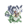 1bsuC 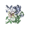 1az0S S: Starting model for refinement C: citing same article ( |
|---|---|
| Similar structure data |


-Links
 PDBj
PDBj


- Assembly
| Deposited unit | 
| ||||||||||
|---|---|---|---|---|---|---|---|---|---|---|---|
| 1 |
| ||||||||||
| Unit cell |
|
-Components
| #1: DNA chain | Mass: 3342.209 Da / Num. of mol.: 2 / Source method: obtained synthetically #2: Protein | Mass: 28559.158 Da / Num. of mol.: 2 Source method: isolated from a genetically manipulated source Source: (gene. exp.) References: UniProt: P04390, type II site-specific deoxyribonuclease #3: Water | ChemComp-HOH / |  Mass: 18.015 Da / Num. of mol.: 236 / Source method: isolated from a natural source / Formula: H2O Mass: 18.015 Da / Num. of mol.: 236 / Source method: isolated from a natural source / Formula: H2O |
|---|
-Experimental details
-Experiment
| Experiment | Method: X-RAY DIFFRACTION / Number of used crystals: 1 |
|---|
- Sample preparation
| Crystal | Density Matthews: 2.17 Å3/Da / Density % sol: 43.23 % | |||||||||||||||||||||||||||||||||||||||||||||||||
|---|---|---|---|---|---|---|---|---|---|---|---|---|---|---|---|---|---|---|---|---|---|---|---|---|---|---|---|---|---|---|---|---|---|---|---|---|---|---|---|---|---|---|---|---|---|---|---|---|---|---|
| Crystal grow | Method: vapor diffusion, hanging drop / pH: 7.5 / Details: pH 7.5, VAPOR DIFFUSION, HANGING DROP | |||||||||||||||||||||||||||||||||||||||||||||||||
| Crystal grow | *PLUS Method: vapor diffusion | |||||||||||||||||||||||||||||||||||||||||||||||||
| Components of the solutions | *PLUS
|
-Data collection
| Diffraction | Mean temperature: 293 K |
|---|---|
| Diffraction source | Source: ROTATING ANODE / Type: RIGAKU RU200 |
| Detector | Type: RIGAKU / Detector: IMAGE PLATE / Date: Aug 15, 1997 / Details: MIRRORS |
| Radiation | Monochromator: MIRRORS / Protocol: SINGLE WAVELENGTH / Monochromatic (M) / Laue (L): M / Scattering type: x-ray |
| Radiation wavelength | Relative weight: 1 |
| Reflection | Resolution: 1.9→20 Å / Num. obs: 37374 / % possible obs: 86 % / Observed criterion σ(I): 1 / Redundancy: 2.3 % / Biso Wilson estimate: 40.7 Å2 / Rmerge(I) obs: 0.056 |
| Reflection | *PLUS % possible obs: 86 % / Num. measured all: 83222 |
| Reflection shell | *PLUS % possible obs: 75.4 % |
- Processing
| Software |
| ||||||||||||||||||||||||||||||||||||||||||||||||||||||||||||
|---|---|---|---|---|---|---|---|---|---|---|---|---|---|---|---|---|---|---|---|---|---|---|---|---|---|---|---|---|---|---|---|---|---|---|---|---|---|---|---|---|---|---|---|---|---|---|---|---|---|---|---|---|---|---|---|---|---|---|---|---|---|
| Refinement | Method to determine structure: OTHER Starting model: PDB 1AZ0 Resolution: 2.15→6 Å / Cross valid method: THROUGHOUT / σ(F): 1 Details: THE AMIMO ACID SIDE CHAINS LISTED IN REMARK 470 APPEAR TO BE PARTIALLY OR COMPLETELY DISORDERED.
| ||||||||||||||||||||||||||||||||||||||||||||||||||||||||||||
| Displacement parameters | Biso mean: 26.5 Å2 | ||||||||||||||||||||||||||||||||||||||||||||||||||||||||||||
| Refine analyze | Luzzati coordinate error obs: 0.25 Å | ||||||||||||||||||||||||||||||||||||||||||||||||||||||||||||
| Refinement step | Cycle: LAST / Resolution: 2.15→6 Å
| ||||||||||||||||||||||||||||||||||||||||||||||||||||||||||||
| Refine LS restraints |
| ||||||||||||||||||||||||||||||||||||||||||||||||||||||||||||
| Xplor file |
| ||||||||||||||||||||||||||||||||||||||||||||||||||||||||||||
| Software | *PLUS Name: X-PLOR / Version: 3.1 / Classification: refinement | ||||||||||||||||||||||||||||||||||||||||||||||||||||||||||||
| Refinement | *PLUS Rfactor obs: 0.2 / Rfactor Rwork: 0.2 | ||||||||||||||||||||||||||||||||||||||||||||||||||||||||||||
| Solvent computation | *PLUS | ||||||||||||||||||||||||||||||||||||||||||||||||||||||||||||
| Displacement parameters | *PLUS |
