 Movie
Movie Controller


+ Open data

- Basic information
| Entry | Database: PDB / ID: 1eon | |||||||||
|---|---|---|---|---|---|---|---|---|---|---|
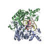
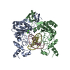
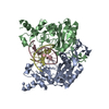
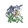
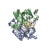
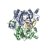
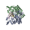
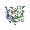
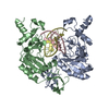
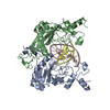
| Title | ECORV BOUND TO 3'-S-PHOSPHOROTHIOLATE DNA AND CA2+ | |||||||||
 Components Components |
| |||||||||
 Keywords Keywords | hydrolase/DNA / protein-nucleic acid recognition / restriction enzyme / DNA analog / metal ion catalysis / hydrolase-DNA COMPLEX | |||||||||
| Function / homology |  Function and homology information Function and homology informationtype II site-specific deoxyribonuclease / type II site-specific deoxyribonuclease activity / DNA restriction-modification system / DNA binding / metal ion binding Similarity search - Function | |||||||||
| Biological species |  | |||||||||
| Method |  X-RAY DIFFRACTION / SYNCHROTRON / Resolution: 1.6 Å X-RAY DIFFRACTION / SYNCHROTRON / Resolution: 1.6 Å | |||||||||
 Authors Authors | Horton, N.C. / Connolly, B.A. / Perona, J.J. | |||||||||
 Citation Citation | Journal: J.Am.Chem.Soc. / Year: 2000 Title: Inhibition of EcoRV Endonuclease by Deoxyribo-3'-S-phosphorothiolates: A High-Resolution X-ray Crystallographic Study Authors: Horton, N.C. / Connolly, B.A. / Perona, J.J. | |||||||||
| History |
|
- Structure visualization
| Structure viewer | Molecule: MolmilJmol/JSmol |
|---|
- Downloads & links
-Download
| PDBx/mmCIF format | 1eon.cif.gz | 128.9 KB | Display | PDBx/mmCIF format |
|---|---|---|---|---|
| PDB format | pdb1eon.ent.gz | 95.5 KB | Display | PDB format |
| PDBx/mmJSON format | 1eon.json.gz | Tree view | PDBx/mmJSON format | |
| Others |  Other downloads Other downloads |
-Validation report
| Arichive directory | https://data.pdbj.org/pub/pdb/validation_reports/eo/1eonftp://data.pdbj.org/pub/pdb/validation_reports/eo/1eon | HTTPS FTP |
|---|
-Related structure data
| Similar structure data |
|---|


-Links
 PDBj
PDBj


- Assembly
| Deposited unit | 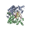
| ||||||||||
|---|---|---|---|---|---|---|---|---|---|---|---|
| 1 |
| ||||||||||
| Unit cell |
| ||||||||||
| Details | The biological assembly is a dimer composed of chains A and B / The biological assembly is a duplex composed of chains C and D |
-Components
-DNA chain , 2 types, 2 molecules CD
| #1: DNA chain | Mass: 3372.301 Da / Num. of mol.: 1 / Source method: obtained synthetically |
|---|---|
| #2: DNA chain | Mass: 3348.276 Da / Num. of mol.: 1 / Source method: obtained synthetically |
-Protein , 1 types, 2 molecules AB
| #3: Protein | Mass: 28690.354 Da / Num. of mol.: 2 Source method: isolated from a genetically manipulated source Source: (gene. exp.) References: UniProt: P04390, type II site-specific deoxyribonuclease |
|---|
-Non-polymers , 3 types, 363 molecules 


| #4: Chemical | ChemComp-CL /  Mass: 35.453 Da / Num. of mol.: 4 / Source method: obtained synthetically / Formula: Cl Mass: 35.453 Da / Num. of mol.: 4 / Source method: obtained synthetically / Formula: Cl#5: Chemical |  Mass: 60.052 Da / Num. of mol.: 2 / Source method: obtained synthetically / Formula: C2H4O2 Mass: 60.052 Da / Num. of mol.: 2 / Source method: obtained synthetically / Formula: C2H4O2#6: Water | ChemComp-HOH / | Mass: 18.015 Da / Num. of mol.: 357 / Source method: isolated from a natural source / Formula: H2O |
|---|
-Experimental details
-Experiment
| Experiment | Method: X-RAY DIFFRACTION / Number of used crystals: 1 |
|---|
- Sample preparation
| Crystal | Density Matthews: 2.03 Å3/Da / Density % sol: 39.55 % | ||||||||||||||||||||||||||||||||||||||||||
|---|---|---|---|---|---|---|---|---|---|---|---|---|---|---|---|---|---|---|---|---|---|---|---|---|---|---|---|---|---|---|---|---|---|---|---|---|---|---|---|---|---|---|---|
| Crystal grow | Temperature: 290 K / Method: vapor diffusion, hanging drop / pH: 7.5 Details: 25% PEG 4000, 0.1 M HEPES, 0.15 M NaCl, 50m M CaCl2, pH 7.5, VAPOR DIFFUSION, HANGING DROP, temperature 290K | ||||||||||||||||||||||||||||||||||||||||||
| Components of the solutions |
| ||||||||||||||||||||||||||||||||||||||||||
| Crystal grow | *PLUS Temperature: 17 ℃ / Method: vapor diffusion | ||||||||||||||||||||||||||||||||||||||||||
| Components of the solutions | *PLUS
|
-Data collection
| Diffraction | Mean temperature: 100 K |
|---|---|
| Diffraction source | Source: SYNCHROTRON / Site: SSRL  / Beamline: BL7-1 / Wavelength: 1.05 / Beamline: BL7-1 / Wavelength: 1.05 |
| Detector | Type: MARRESEARCH / Detector: IMAGE PLATE / Date: Jan 20, 1999 |
| Radiation | Protocol: SINGLE WAVELENGTH / Monochromatic (M) / Laue (L): M / Scattering type: x-ray |
| Radiation wavelength | Wavelength: 1.05 Å / Relative weight: 1 |
| Reflection | Resolution: 1.58→20 Å / Num. all: 65611 / Num. obs: 65611 / % possible obs: 94.7 % / Observed criterion σ(I): -3 / Redundancy: 1.96 % / Biso Wilson estimate: 23.1 Å2 / Rmerge(I) obs: 0.037 / Net I/σ(I): 8.9 |
| Reflection shell | Resolution: 1.58→1.63 Å / Redundancy: 2 % / Rmerge(I) obs: 0.32 / Num. unique all: 5936 / % possible all: 92.8 |
| Reflection | *PLUS Num. measured all: 128582 |
| Reflection shell | *PLUS % possible obs: 92.8 % / Rmerge(I) obs: 0.324 / Mean I/σ(I) obs: 3 |
- Processing
| Software |
| |||||||||||||||||||||||||
|---|---|---|---|---|---|---|---|---|---|---|---|---|---|---|---|---|---|---|---|---|---|---|---|---|---|---|
| Refinement | Resolution: 1.6→4.8 Å / Cross valid method: THROUGHOUT / σ(F): 2 / σ(I): 1 / Stereochemistry target values: X-Plor
| |||||||||||||||||||||||||
| Refinement step | Cycle: LAST / Resolution: 1.6→4.8 Å
| |||||||||||||||||||||||||
| Refine LS restraints |
| |||||||||||||||||||||||||
| Software | *PLUS Name: X-PLOR / Classification: refinement | |||||||||||||||||||||||||
| Refinement | *PLUS σ(F): 2 / % reflection Rfree: 2 % / Rfactor obs: 0.229 | |||||||||||||||||||||||||
| Solvent computation | *PLUS | |||||||||||||||||||||||||
| Displacement parameters | *PLUS Biso mean: 23.1 Å2 | |||||||||||||||||||||||||
| Refine LS restraints | *PLUS Type: x_angle_deg / Dev ideal: 2.4 |
