 Movie
Movie Controller


+ Open data

- Basic information
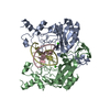
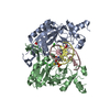
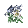
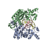
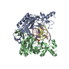
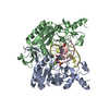
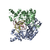
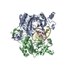
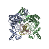
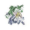
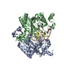
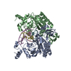
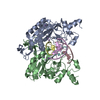
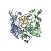
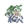
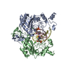
| Entry | Database: PDB / ID: 2b0e | ||||||
|---|---|---|---|---|---|---|---|
| Title | EcoRV Restriction Endonuclease/GAAUTC/Ca2+ | ||||||
 Components Components |
| ||||||
 Keywords Keywords | HYDROLASE/DNA / protein-nucleic acid recognition / indirect readout / restriction enzyme / substrate specificity / noncognate / HYDROLASE-DNA COMPLEX | ||||||
| Function / homology |  Function and homology information Function and homology informationtype II site-specific deoxyribonuclease / type II site-specific deoxyribonuclease activity / DNA restriction-modification system / DNA binding / metal ion binding Similarity search - Function | ||||||
| Biological species |  | ||||||
| Method |  X-RAY DIFFRACTION / FOURIER SYNTHESIS / Resolution: 1.9 Å X-RAY DIFFRACTION / FOURIER SYNTHESIS / Resolution: 1.9 Å | ||||||
 Authors Authors | Hiller, D.A. / Rodriguez, A.M. / Perona, J.J. | ||||||
 Citation Citation | Journal: J.Mol.Biol. / Year: 2005 Title: Non-cognate Enzyme-DNA Complex: Structural and Kinetic Analysis of EcoRV Endonuclease Bound to the EcoRI Recognition Site GAATTC Authors: Hiller, D.A. / Rodriguez, A.M. / Perona, J.J. #1: Journal: Nat.Struct.Biol. / Year: 1999Title: Structural and energetic origins of indirect readout in site-specific DNA cleavage by a restriction endonuclease Authors: Martin, A.M. / Sam, M.D. / Reich, N.O. / Perona, J.J. #2: Journal: Biochemistry / Year: 2003Title: Simultaneous DNA binding and bending by EcoRV endonuclease observed by real-time fluorescence Authors: Hiller, D.A. / Fogg, J.M. / Martin, A.M. / Beechem, J.M. / Reich, N.O. / Perona, J.J. #3: Journal: Biochemistry / Year: 2004Title: DNA cleavage by EcoRV endonuclease: two metal ions in three metal ion binding sites Authors: Horton, N.C. / Perona, J.J. | ||||||
| History |
|
- Structure visualization
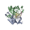
| Structure viewer | Molecule: MolmilJmol/JSmol |
|---|
- Downloads & links
-Download
| PDBx/mmCIF format | 2b0e.cif.gz | 125.8 KB | Display | PDBx/mmCIF format |
|---|---|---|---|---|
| PDB format | pdb2b0e.ent.gz | 92.4 KB | Display | PDB format |
| PDBx/mmJSON format | 2b0e.json.gz | Tree view | PDBx/mmJSON format | |
| Others |  Other downloads Other downloads |
-Validation report
| Arichive directory | https://data.pdbj.org/pub/pdb/validation_reports/b0/2b0eftp://data.pdbj.org/pub/pdb/validation_reports/b0/2b0e | HTTPS FTP |
|---|
-Related structure data


-Links
 PDBj
PDBj


- Assembly
| Deposited unit | 
| ||||||||
|---|---|---|---|---|---|---|---|---|---|
| 1 |
| ||||||||
| Unit cell |
|
-Components
| #1: DNA chain | Mass: 3342.209 Da / Num. of mol.: 2 / Source method: obtained synthetically #2: Protein | Mass: 28690.354 Da / Num. of mol.: 2 Source method: isolated from a genetically manipulated source Source: (gene. exp.) References: UniProt: P04390, type II site-specific deoxyribonuclease #3: Chemical |   Mass: 40.078 Da / Num. of mol.: 2 / Source method: obtained synthetically / Formula: Ca Mass: 40.078 Da / Num. of mol.: 2 / Source method: obtained synthetically / Formula: Ca#4: Water | ChemComp-HOH / |  Mass: 18.015 Da / Num. of mol.: 253 / Source method: isolated from a natural source / Formula: H2O Mass: 18.015 Da / Num. of mol.: 253 / Source method: isolated from a natural source / Formula: H2O |
|---|
-Experimental details
-Experiment
| Experiment | Method: X-RAY DIFFRACTION / Number of used crystals: 1 |
|---|
- Sample preparation
| Crystal | Density Matthews: 1.96 Å3/Da / Density % sol: 37.2 % | ||||||||||||||||||||||||||||||||||||||||||||
|---|---|---|---|---|---|---|---|---|---|---|---|---|---|---|---|---|---|---|---|---|---|---|---|---|---|---|---|---|---|---|---|---|---|---|---|---|---|---|---|---|---|---|---|---|---|
| Crystal grow | Temperature: 297 K / Method: vapor diffusion, hanging drop / pH: 7.5 Details: PEG 4K, Hepes, NaCl, CaCl2, pH 7.5, vapor diffusion, hanging drop, temperature 297K | ||||||||||||||||||||||||||||||||||||||||||||
| Components of the solutions |
|
-Data collection
| Diffraction | Mean temperature: 100 K |
|---|---|
| Diffraction source | Source: ROTATING ANODE / Type: RIGAKU / Wavelength: 1.54 Å |
| Detector | Type: RIGAKU RAXIS II / Detector: IMAGE PLATE / Date: Jun 1, 2000 |
| Radiation | Protocol: SINGLE WAVELENGTH / Scattering type: x-ray |
| Radiation wavelength | Wavelength: 1.54 Å / Relative weight: 1 |
| Reflection | Resolution: 1.9→19.3 Å / Num. obs: 69729 / % possible obs: 90 % / Net I/σ(I): 10.3 |
| Reflection shell | Resolution: 1.9→2 Å / Mean I/σ(I) obs: 2.9 / % possible all: 88.1 |
- Processing
| Software |
| ||||||||||||||||||||||||||||
|---|---|---|---|---|---|---|---|---|---|---|---|---|---|---|---|---|---|---|---|---|---|---|---|---|---|---|---|---|---|
| Refinement | Method to determine structure: FOURIER SYNTHESIS / Resolution: 1.9→6 Å /
| ||||||||||||||||||||||||||||
| Displacement parameters | Biso mean: 19.625 Å2 | ||||||||||||||||||||||||||||
| Refinement step | Cycle: LAST / Resolution: 1.9→6 Å
| ||||||||||||||||||||||||||||
| Refine LS restraints |
|
