 Movie
Movie Controller


+ Open data

- Basic information
| Entry | Database: PDB / ID: 1bgb | ||||||
|---|---|---|---|---|---|---|---|
| Title | ECORV ENDONUCLEASE COMPLEX WITH 5'-CGGGATATCCC DNA | ||||||
 Components Components |
| ||||||
 Keywords Keywords | HYDROLASE/DNA / COMPLEX (ENDONUCLEASE-DNA) / HYDROLASE-DNA COMPLEX | ||||||
| Function / homology |  Function and homology information Function and homology informationtype II site-specific deoxyribonuclease / type II site-specific deoxyribonuclease activity / DNA restriction-modification system / DNA binding / metal ion binding Similarity search - Function | ||||||
| Biological species |  | ||||||
| Method |  X-RAY DIFFRACTION / DIFFERENCE FOURIER / Resolution: 2 Å X-RAY DIFFRACTION / DIFFERENCE FOURIER / Resolution: 2 Å | ||||||
 Authors Authors | Perona, J. / Horton, N.C. | ||||||
 Citation Citation | Journal: J.Biol.Chem. / Year: 1998 Title: Recognition of flanking DNA sequences by EcoRV endonuclease involves alternative patterns of water-mediated contacts. Authors: Horton, N.C. / Perona, J.J. #1: Journal: J.Mol.Biol. / Year: 1998Title: Role of Protein-Induced Bending in the Specificity of DNA Recognition-Crystal Structure of EcoRV Endonuclease Complexed with D(Aaagat) + D(Atctt) Authors: Horton, N.C. / Perona, J.J. #2: Journal: J.Mol.Biol. / Year: 1997Title: Conformational Transitions and Structural Deformability of EcoRV Endonuclease Revealed by Crystallographic Analysis Authors: Perona, J.J. / Martin, A.M. | ||||||
| History |
|
- Structure visualization
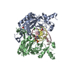
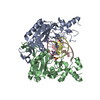
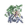
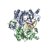
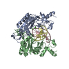
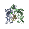
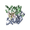
| Structure viewer | Molecule: MolmilJmol/JSmol |
|---|
- Downloads & links
-Download
| PDBx/mmCIF format | 1bgb.cif.gz | 120.6 KB | Display | PDBx/mmCIF format |
|---|---|---|---|---|
| PDB format | pdb1bgb.ent.gz | 89.3 KB | Display | PDB format |
| PDBx/mmJSON format | 1bgb.json.gz | Tree view | PDBx/mmJSON format | |
| Others |  Other downloads Other downloads |
-Validation report
| Arichive directory | https://data.pdbj.org/pub/pdb/validation_reports/bg/1bgbftp://data.pdbj.org/pub/pdb/validation_reports/bg/1bgb | HTTPS FTP |
|---|
-Related structure data
| Similar structure data |
|---|


-Links
 PDBj
PDBj


- Assembly
| Deposited unit | 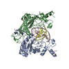
| ||||||||||
|---|---|---|---|---|---|---|---|---|---|---|---|
| 1 |
| ||||||||||
| Unit cell |
|
-Components
| #1: DNA chain | Mass: 3334.186 Da / Num. of mol.: 2 / Source method: obtained synthetically #2: Protein | Mass: 28559.158 Da / Num. of mol.: 2 Source method: isolated from a genetically manipulated source Source: (gene. exp.) References: UniProt: P04390, type II site-specific deoxyribonuclease #3: Water | ChemComp-HOH / |  Mass: 18.015 Da / Num. of mol.: 236 / Source method: isolated from a natural source / Formula: H2O Mass: 18.015 Da / Num. of mol.: 236 / Source method: isolated from a natural source / Formula: H2O |
|---|
-Experimental details
-Experiment
| Experiment | Method: X-RAY DIFFRACTION / Number of used crystals: 2 |
|---|
- Sample preparation
| Crystal | Density Matthews: 2.18 Å3/Da / Density % sol: 43.63 % | ||||||||||||||||||||||||||||||||||||||||||
|---|---|---|---|---|---|---|---|---|---|---|---|---|---|---|---|---|---|---|---|---|---|---|---|---|---|---|---|---|---|---|---|---|---|---|---|---|---|---|---|---|---|---|---|
| Crystal grow | Method: vapor diffusion, hanging drop / pH: 6.5 Details: CRYSTALLIZED FROM 15% PEG 4K, 100 MM IMIDAZOLE, (PH 6.5), 150 MM NACL., VAPOR DIFFUSION, HANGING DROP | ||||||||||||||||||||||||||||||||||||||||||
| Components of the solutions |
| ||||||||||||||||||||||||||||||||||||||||||
| Crystal grow | *PLUS Method: vapor diffusion | ||||||||||||||||||||||||||||||||||||||||||
| Components of the solutions | *PLUS
|
-Data collection
| Diffraction | Mean temperature: 293 K |
|---|---|
| Diffraction source | Source: ROTATING ANODE / Type: RIGAKU |
| Detector | Type: RIGAKU RAXIS IIC / Detector: IMAGE PLATE / Date: Aug 15, 1997 / Details: MIRRORS |
| Radiation | Monochromator: NI FILTER / Monochromatic (M) / Laue (L): M / Scattering type: x-ray |
| Radiation wavelength | Relative weight: 1 |
| Reflection | Resolution: 2.1→20 Å / Num. obs: 25665 / % possible obs: 85 % / Observed criterion σ(I): -3 / Redundancy: 2.2 % / Biso Wilson estimate: 28.2 Å2 / Rmerge(I) obs: 0.053 / Rsym value: 0.091 / Net I/σ(I): 10 |
| Reflection shell | Resolution: 2.1→2.2 Å / Redundancy: 1.9 % / Rmerge(I) obs: 0.077 / Mean I/σ(I) obs: 2.8 / Rsym value: 0.28 / % possible all: 71 |
- Processing
| Software |
| ||||||||||||||||||||||||||||||||||||||||||||||||||||||||||||
|---|---|---|---|---|---|---|---|---|---|---|---|---|---|---|---|---|---|---|---|---|---|---|---|---|---|---|---|---|---|---|---|---|---|---|---|---|---|---|---|---|---|---|---|---|---|---|---|---|---|---|---|---|---|---|---|---|---|---|---|---|---|
| Refinement | Method to determine structure: DIFFERENCE FOURIER / Resolution: 2→6 Å / Rfactor Rfree error: 0.017 / Data cutoff high absF: 100000 / Data cutoff low absF: 0.1 / Isotropic thermal model: RESTRAINED / Cross valid method: THROUGHOUT / σ(F): 1
| ||||||||||||||||||||||||||||||||||||||||||||||||||||||||||||
| Displacement parameters | Biso mean: 27.7 Å2 | ||||||||||||||||||||||||||||||||||||||||||||||||||||||||||||
| Refine analyze |
| ||||||||||||||||||||||||||||||||||||||||||||||||||||||||||||
| Refinement step | Cycle: LAST / Resolution: 2→6 Å
| ||||||||||||||||||||||||||||||||||||||||||||||||||||||||||||
| Refine LS restraints |
| ||||||||||||||||||||||||||||||||||||||||||||||||||||||||||||
| LS refinement shell | Resolution: 2.1→2.2 Å / Rfactor Rfree error: 0.042 / Total num. of bins used: 8
| ||||||||||||||||||||||||||||||||||||||||||||||||||||||||||||
| Xplor file |
| ||||||||||||||||||||||||||||||||||||||||||||||||||||||||||||
| Software | *PLUS Name: X-PLOR / Classification: refinement | ||||||||||||||||||||||||||||||||||||||||||||||||||||||||||||
| Refine LS restraints | *PLUS
|
